Phyllodes tumor is a rare breast malignancy of stromal tissue. They are benign, borderline and malignant as per WHO criteria. The mainstay of treatment is surgery. These tumors mimic benign conditions and are tricky to diagnose. We are here reporting a rare case of Phyllodes tumor left breast in a 45 yr old female. She was treated with surgery for borderline Phyllodes tumor and owing to large size more than 10 cm and positive margins received adjuvant radiation therapy.
Key findings:
Key findings highlight Phyllodes tumor, a rare breast malignancy with benign, borderline, and malignant variants. Treatment primarily involves surgery, but challenges exist in diagnosis due to its benign mimicry. The case report details a 45-year-old female with a borderline Phyllodes tumor, emphasizing the use of adjuvant radiation therapy for large tumors with positive margins.
What is known and what is new?
The abstract outlines Phyllodes tumor as a rare breast malignancy with benign, borderline, and malignant variants, typically treated with surgery. It emphasizes the diagnostic challenge due to its mimicry of benign conditions. The case report introduces a novel aspect, highlighting the use of adjuvant radiation therapy for a large borderline Phyllodes tumor with positive margins.
What is the implication, and what should change now?
The case highlights the challenges in diagnosing and managing Phyllodes tumors, emphasizing the need for heightened clinical suspicion and comprehensive evaluation of breast masses. The utilization of adjuvant radiation therapy for cases with large size and positive margins suggests the importance of personalized treatment strategies to optimize outcomes and minimize recurrence risks.
Phyllodes tumor is a rare tumor of the breast accounting for less than 1% of all breast malignancies and has an incidence of about 2.1 per million[1]. These tumors start in the connective tissue (stroma) of the breast with mixed gelatinous cystic and solid areas [2]. They commonly affect women during the 4th or 5th decade of life [2]. Males are rarely affected. Phyllodes tumors are benign, borderline and malignant depending on cytologic features given by WHO. The mainstay treatment of phyllodes tumor is surgery, breast conservative or total mastectomy [2]. Tumors larger than 10 cm in size are giant Phyllodes tumors and are difficult to manage surgically as it is difficult to achieve negative margins. The borderline Phyllodes tumors have the greatest tendency for local recurrence [3]. Local recurrence depends on several factors like giant tumor size, positive margins among them [4]. Radiotherapy after breast surgery may significantly reduce local recurrence [5]. However, due to rarity of disease there are a limited number of trials for the role of radiotherapy in giant Phyllodes tumor.
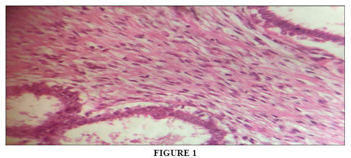
A 45 year old patient presented to us with awareness of a lump in left breast for about 2 years. She had a long clinical history of gradually progressive without any associated pain. From the last 3 months there is a history of ulceration and bleeding from the lump. There is a history of severe pain associated with it. For these complaints she visited the local hospital where biopsy was done suggestive of Phyllodes tumor left breast and then was referred to our centre. On examination of the patient there was a mass in the left outer quadrant of breast measuring about 10 x 12 cm with overlying skin ulceration and bleeding. There was left axillary lymphadenopathy about 1x1cm single node. Further work up was done. Biochemical parameters were within normal range. MRI breast was suggestive of 12.5x10.5 x 13.9 cm lobulated mass involving whole breast and overlying skin and bulging exophytically out. A total mastectomy was done at the surgical department. On gross pathological examination there was a 12.5x10x13.5 cm large gray brown mass with focal clefts and nodular appearance involving overlying skin with distorted nipple. Growth was reaching up to a deep resection margin. On microscopic examination there were cleft like spaces, stromal overgrowth with areas revealing mildly pleomorphic cells having increased nucleo cytoplasmic ratio, inconspicuous nucleoli and eosinophilic cytoplasm. Areas of pseudo angiomatous pattern of stroma and myxoid changes were present. There was increased mitosis with focal epithelial hyperplasia in a leaf pattern.
Clusters of benign acini and hyalinization were seen [Figure 1]. A final diagnosis of Phyllodes tumor was made. Tumor was reaching within 1 mm of deep resection margin. Circumferential margin was involved. Patient was then planned for radiation therapy to the left chest wall. A dose of 40 Gray in 15 fractions over 3 weeks was delivered. Patient was examined weekly for radiation toxicity. There was grade 1 skin toxicity. Patient was advised regular physiotherapy. Patient has now completed 1 year of close follow up and is disease free.
Phyllodes tumor is a rare breast tumor which starts in the stromal tissue [1,2]. It is a well circumscribed tumor which grows rapidly compressing the surrounding breast parenchyma creating a false capsule through which it further spreads. It is usually painless. It affects the left breast more often than the right one with female predominance [6]. Our patient's left breast was affected. There was a large tumor more than 10 cm in size with ulceration over skin.
Phyllodes tumor grows rapidly and mimics fibroadenoma making it difficult to diagnose. Mammography and breast ultrasound cannot differentiate Phyllodes from fibroadenoma [7]. A biopsy, incisional or excisional or core cut is most reliable for the diagnosis. Phyllodes tumors are classified by WHO into benign, borderline or malignant based on degree of stromal atypia, mitotic activity, and stromal overgrowth. Borderline tumors are most recurrent [3].
Treatment of choice for borderline Phyllodes tumor is complete surgical resection, however if tumor size is large there is difficulty in achieving negative margins as they penetrate into surrounding healthy tissue [8]. It has been seen that positive margin, tumor size, high mitotic count, stromal overgrowth and necrosis are factors that increase chances of local recurrence. The chances increase by four times for tumor size >10cm and by fivefold if tumor margin is less than 1 cm [9]. In our patient there were high chances of tumor recurrence as both factors i.e. large tumor size >10cm and margin involvement were present. Because of this she received local radiation therapy as adjuvant, as radiation therapy has a role in preventing local recurrence in this condition.
A quick and proper diagnosis with meticulous treatment is required for treatment of Phyllodes tumor and to prevent its recurrence. This is difficult to achieve as it mimics benign conditions.
Phyllodes tumor of breast is a rare malignancy. It is difficult to diagnose and is highly recurrent, especially the borderline subtype. A quick accurate diagnosis supported preferably by biopsy is required. Surgery is the mainstay of treatment with adjuvant radiation in special conditions. More studies with the role of adjuvant radiation therapy are warranted in future.
Funding: No funding sources.
Conflict of interest: None declared.
Ethical approval: The study was approved by the Institutional Ethics Committee of Shri Lal Bahadur Shastri Medical College.