Contents
Download PDF
pdf Download XML
2927 Views
321 Downloads
Share this article
Research Article | Volume 2 Issue 2 (July-Dec, 2021) | Pages 1 - 6
A Cross Sectional Study to Evaluate the Impact of Malignancy on Breast Density
 ,
1
Department of General Surgery, Indira Gandhi Medical College Shimla, 171001, H.P, India
Under a Creative Commons license
Open Access
Received
July 5, 2021
Revised
Aug. 14, 2021
Accepted
Aug. 26, 2021
Published
Sept. 10, 2021
Abstract

Background: Mammographic density has been shown to be a strong independent predictor of breast cancer and a causative factor in reducing the sensitivity of mammography. The risk of breast cancer related to changes in breast density over time, including its regression and persistence, remains controversial. The present study was done to evaluate the impact of malignancy on Breast Density. Material and Methods: Present study was conducted in the Department of General Surgery and Radio diagnosis IGMC Shimla from 1st July 2018 to 30 June 2019. A total of 60 were included and evaluated in the present study. These patients were divided into two groups. Group (A) included 30 patients with biopsy proven carcinoma of the breast while Group (B) included 30 patients with benign breast lesions or pain breast. Results: The mean age of patients presenting in Group A was 55.4±10.9 years and the mean age of the patients presenting in Group B was 54±9.84years respectively. In Group A of 30 cancer patients, majority of patients i.e 21patients (70%) had breast density of Type B. In Group B also, majority 20 patients (66.7%) had breast density Type B. In Group A out of 30 cancer patients, 10patients (i.e.33.3%) were BIRADS 4,14 patients (46.7%) were BIRADS 5 and 6 patients (20%) were BIRADS 6. In Group B out of 30 patients, 6 patients (i.e.20%) were BIRADS 1,12 patients (40%) were BIRADS 2 and 12 patients (40%) were BIRADS 3. In Group A of 30 cancer patients ,27 patients were diagnosed with duct cell carcinoma (90%) while 2 patient were reported as lobular carcinoma (6.7%) and 1 patients was diagnosed with tubular carcinoma(3.3%) . In Group B out of 30 patients, 2 patients (6.7%) were diagnosed as acute mastitis, 19 patients (63.3%) were diagnosed as Fibro-adenoids, 3patients (10%) were diagnosed as Fibro-adenoma. Conclusion: Maximum patients in this study were in the age group of 40-60 year. Most breast cancer patients had higher BIRADS 5 and 6 , had breast density of Type B and majority of were reported as Duct Cell Carcinoma. Most of the benign breast disease patients had BIRADS2 and 3, had breast density of Type B and majority of were reported as Fibro-adenoids.

Keywords
INTRODUCTION

Breast density reflects the amount of fibrous and glandular tissue in a woman’s breasts compared with the amount of fatty tissue in the breasts. Breast density is seen only on mammograms. Breast density isn’t based on how your breasts feel, and it’s not related to breast size or firmness. Women who have dense breast tissue have a higher risk of breast cancer compared to women with less dense breast tissue. The denser your breasts are, the higher your risk [1,2].


However, patients don’t necessarily have a high risk of breast cancer just because she has dense breasts. Breast density has to be considered along with other risk factors, such as age, family history, and any history of breast biopsies showing atypical cells or other changes that increase cancer risk [1,2]. There is no consensus on the most useful measure of breast composition in risk prediction, risk management and surveillance decisions. One meta-analysis found that absolute rather than proportional estimates of breast density are more strongly predictive of risk, whereas another found the opposite [3] Numerous studies have shown that breast density, as assessed through mammography, is an important breast cancer risk factor. Relative to the lowest classification of breast density (fatty tissue), women with the highest classification (extreme density) may have a 2-to-6-fold increased risk of breast cancer [4]. There is paucity of studies on relation of breast density with breast 


cancer. Against this background, resent study was done to evaluate the impact of malignancy on Breast Density.


Aims and objectives

To evaluate the impact of malignancy on Breast Density

MATERIALS AND METHODS

Study area 

Present study was conducted in the Department of General Surgery and Radio diagnosis IGMC Shimla over a period of one year. 


Study period 

From 1st July 2018 to 30 June 2019. 


Study Participants

Patients presenting with the symptoms of breast lump, breast pain, swelling and nipple discharge in the Department of Surgery were included in the present study. 


Sample size 

A total of 60 were included and evaluated in the present study. 

These patients were divided into two groups: 

Group (A) included 30 patients with biopsy proven carcinoma of the breast. 

Group (B) included 30 patients with benign breast lesions or pain breast. 


Inclusion Criteria 

Patients above 40 years with complaints of lump breast, breast pain, swelling, discharge from the nipple, or biopsy proven carcinoma of the breast were included in the present study.


Exclusion Criteria 


  • Patients less than 40 years. 

  • Patients who had undergone Breast surgery for Benign or Malignant pathology. 

  • Patients who have previously received Radio Therapy 


Method of Data Collection

Informed consent was taken from all the patients. In every case detailed history was taken and a thorough clinical examination was done. The following investigations were done in every case. 


  • Complete haemogram 

  • Renal Function Test 

  • Liver Function Test 

  • Chest X-ray 

  • Mammography 


Mammography 

Bilateral Mammography was performed in all the patients of Group A and Group B over 40 yrs of age with standard planes of imaging i.e cranio-caudal (CC) and mediolateral oblique (MLO) view. All the mammograms were done with Fujifilm Mammographic Unit installed at IGMC Shimla by an experienced radiologist. 

                In cranio-caudal view the x-ray films were placed underneath the breast. The breast was held against the image receptor with compression. Mediolateral view was obtained with the x-rays film held between and parallel to the pectoralis major and latssimus dorsi muscles. 

                With comparative study of mammograms of the two breasts, the mammographic features were recorded. Breast density was determined by using Breast Imaging-Reporting and Data System (BI-RADS). 


  • Additional imaging or prior examinations 

  • Negative 

  • Benign 

  • Probably Benign 

  • Suspicious 

  • Highly Suggestive of Malignancy 

  • Biopsy proven 

  •  

Breast density measured by BI-RADS was further classified into 4 Types;


  • Type A (0%-25%): mostly fatty breast

  • Type B (25%-50%): fibro glandular breast

  • Type C (50%-75%): heterogeneously dense breast, and 

  • Type D (75%-100%): extremely dense breast on the basis of breast composition


Patients were subjected as per BIRADS guidelines for FNAC/ 34 


Trucut Biopsy

Biopsy/FNAC obtained in relevant cases was sent for histological examination where it was analyzed by a faculty member of Department of Pathology. The histo-pathological report was then subsequently obtained and duly noted.


Statistical Analysis 

Results obtained were analyzed statistically. Data for the above mentioned result was compiled, tabulated, statistically analyzed using standard statistical methods. The relevance of result in the light statistical analysis was displayed and discussed.

RESULTS

Results and Observation

The present study was conducted in the Department of Surgery, IGMC, Shimla over one year period from 1st July 2018 to 30 June 2019.The present study comprised of 60 patients above 40 years with breast complaints after taking informed consents. Patients were divided into two groups of 30 patients each. Group A included 30 patients with malignant lesion in the breast, while Group B included 30 patients with benign cause of breast lump, pain and those patients who were subjected for screening mammography. In every case detailed history was taken and thorough clinical examination was done. Ultrasonography, Mammography and trucut biopsy/FNAC was carried out in Group A while Screening Mammography was carried out in Group B. The mean age of patients 


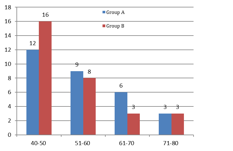
presenting in Group A was 55.4±10.9 years and the mean age of the patients presenting in Group B was 54±9.84years respectively. In group A, maximum 12 patients were in the age group of 40-50 years followed by 9 patients in age group of 51-60 years. Also in group B, maximum 16 patients were in the age group of 40-50 years followed by 8 in age group of 51-60 years (Table 1) (Figur 1). 


Figure1: Age group of breast cancer patients


Table 1: Age group of breast cancer patients 

Age Groups (In Years) Group A Group B 
40-50 12 16 
51-60 
61-70 
71-80 
Total (No .Of Patients) 30 30 


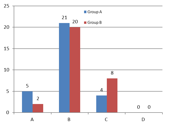
Figure 2: Breast density in patients


Table 2: Breast density in patients

  Group A Group B
Breast Density No. of Patients Percentage No. of Patients Percentage 
16.7% 6.6% 
21 70% 20 66.7% 
13.3% 26.7% 
0% 0% 
Total 30 100% 30 100% 


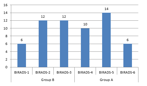
Figure 3: BIRADS in patients


Table-3: BIRADS in patients

  Group A Group B
BIRADS No. of Patients Percentage No. of Patients Percentage 
100%20% 
200%12 40% 
300%12 40% 
10 33.3% 00%
14 46.7% 00%
20% 00%
Total 30 100% 30 100% 


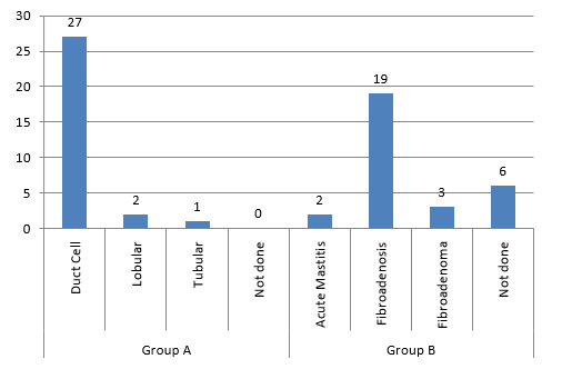
Figure 4: Diagnosis in both groups 


Table 4: Diagnosis in both groups as per FNAC

 Group A Group B
Number of Patients Biopsy Result Percentage Number of Patients FNAC Result Percentage 
27 Duct Cell 90% Acute Mastitis 6.7% 
Lobular 6.7% 19 Fibroadenosis 63.3% 
Tubular 3.3% Fibroadenoma 10% 
0Not done 0%Not done 20% 
30Total 100%30Total 100%


Table-5:

Density Group A Group B Z Score P Value 
1.21 0.23 
21 20 0.28 0.78 
1.29 0.19 
Total 30 30 


No significant family history was present in any patient of either group (A, B). In Group A of 30 cancer patients, 5 patients (16.7%) had breast density of Type A i.e. mostly fatty, while majority of patients i.e 21patients (70%) had breast density of Type B i.e. scattered fibro glandular density and 4 patients (13.3%) had breast density of Type C i.e. consistently dense whereas no patient had Type D density. In Group B, 2 patients (6.6%) had breast density Type A i.e. mostly fatty, while majority 20 patients (66.7%) had breast density Type B i.e. scattered fibro glandular density while in 8 patients (26.7%) the breast density was Type C i.e. consistently dense whereas no patient was of Type D density in this group also (Table 2) (Figure 2).


In Group A out of 30 cancer patients, 10patients (i.e.33.3%) were BIRADS 4, 14 patients (46.7%) were BIRADS 5 and 6 patients (20%) were BIRADS 6. All of the 30 patients were subjected for Trucut Biopsy. In Group B out of 30 patients, 6 patients (i.e.20%) were BIRADS 1, 12 patients (40%) were BIRADS 2 and 12 patients (40%) were BIRADS 3. All Out of 30 patients 24 patients were subjected for FNAC whereas 6 patients with BIRADS 1 were not subjected for FNAC. (Table -3) (Figure 3).


In Group A of 30 cancer patients Trucut biopsy was done in all and 27 patients were diagnosed with duct cell carcinoma (90%) while 2 patient were reported as lobular carcinoma (6.7%) and 1 patients was diagnosed with tubular carcinoma(3.3%). In Group B out of 30 patients, 2 patients (6.7%) were diagnosed as acute mastitis, 19 patients (63.3%) were diagnosed as Fibro-adenoids, 3 patients (10%) were diagnosed as Fibro-adenoma, and 6 patients (20%) were not subjected for FNAC due to BIRADS 1on mammography (Table 4) (Figure 4).


On comparing the proportion of Group A and Group B with density A it was found to be statistically non-significant (z score 1.21 and p value 0.23). The proportion of density B was found to be statistically non-significant (score 0.28 and p value 0.78) similarly the proportion of density C was also found to be statistically non-significant (z score -1.29 and p value 0.19.


DISCUSSION

Breast density is a measurement of the ratio between radio dense epithelium and stroma to radiolucent fatty tissue. Increased breast density as identified on mammography is also known to decrease the diagnostic sensitivity of the examination, which is of great concern to women at increased risk for breast cancer. However, few studies have characterized the incidence of heterogeneously dense or extremely dense tissue in breast cancer patients.


Breast density is a mammographic finding not related to the perceived density of breast tissue on palpation. It is possible to predict with considerable accuracy which women will develop breast cancer and equally important, those who are less likely to develop it based solely on the parenchymal pattern as seen by mammography.


The mean age of patients presenting in Group A was 55.4 ± 10.9 years (ranging from 40 years to 80 years) and the mean age of the patients presenting in Group B was 54±9.84 years (ranging from 40 years to 80 years) respectively. Maximum patients in this study were in the age group of 40-60year with 45(75%) out of 60 patients in both groups. Both the groups were comparable in terms of mean age of presentation. 


In the study by Checka et al5(2012) a total of 7007 patients were screened by mammography and the median age of their cohort was found to be 57 years which is comparable to the mean age of presentation of the present study groups. In that study 3867(55.18%) patients belonged to age group between 40-60 with remaining patients being younger or older to this group. Thus the majority of patients presented in the same age group as detected in the present study. 


In another study by Lim Se-Eun et al. 6 (2019) the average age for presentation for mammography cases was 46.86 years for study and 48.51 years for controls respectively. This too is comparable with the results of the present study showing maximum females seek hospital care for their ailments in the middle age group whether pathology is benign or malignant.


In present study with Group A of 30 cancer patients 10patients (i.e.33.3%) were BIRADS 4, 14 patients (46.7%) were BIRADS 5 and 6 patients (20%) were BIRADS 6 with all patients being subjected for Trucut Biopsy. Similarly In Group B, 6patients (20%) were BIRADS 1, 12 patients (40%) were BIRADS 2 and 12 patients (40%) were BIRADS 3. The density values were almost equally distributed between the lower (BIRADS 1 and 2) (60%) and upper (BI-RADS 3 and 4) (73.3%) groupings. BIRADS rating was a strong predictor of presence of malignant pathology in breast as all 30(100%) patients with BIRADS scoring of 4 and above had carcinoma on histopathological correlation while those with lower BIRADS scoring had benign pathologies. Mammography thus appears to be a highly sensitive modality in detect in pathologies of carcinoma breast even in the present scenario with advent of modern imaging techniques such as MRI and Ultrasound. 


In study by Li.T, Li. J,Dai.M et al [7], Women in their study predominately experienced scattered fibro glandular (37.64%) and heterogeneous MD (49.89%); however, the density values were almost equally distributed between the lower (BIRADS 1 and 2) (47.57%) and upper (BI-RADS 3 and 4) (52.43%) groupings and the result was comparable to those of the present study.


In present study of 30 cancer patients, 5 patients (16.7% ) had breast density of Type A i.e. mostly fatty, while majority of patients i.e 21patients (70%) had breast density of Type B i.e. scattered fibro glandular density and 4 patients (13.3%) had breast density of Type C i.e. consistently dense whereas no patient had Type D density. In Group B, of our study, 2 patients (6.6% ) had breast density Type A i.e. mostly fatty , while majority 20 patients(66.7%) had breast density Type B i.e. scattered fibro glandular density while in 8 patients (26.7%) the breast density was Type C i.e. consistently dense whereas no patient was of Type D density in this group .Thus the predominant appearance of breasts on mammogram in both the groups was either fibro glandular(Type B) or consistently dense(Type C) with no statistical difference between the two groups in regards to breast density. 


Even though it is expected that the Group A patients should report with higher breast density as compared to the Group B, however as a multitude of factors are involved in the causation of carcinoma breast and no single isolated factor can be completely responsible for the pathology it is difficult to make generalizations on basis of a single result of a small sample size study. A large sample size randomized trial might provide more insight into the same. 


In a study by Checka et al5 (2012) There was a total of 558 women (8%) with Type A density (predominantly fatty) on mammography, 2570 (37%) with Type B (scattered fibro glandular elements) 3234 (46%) with Type C (heterogeneously dense), and 645 (9%) with Type D (extremely dense) showing predominance of Type B(68.3%) and C(20%) as already mentioned in the present study. Thus the predominant appearance of breasts on mammogram in both the groups was Type B with no statistical difference between the two groups in regards to breast density. 


Limitations 

The limitations of this study are as follows: 

The existence of selection bias should be considered. The study population was selected from single institute with small sample size and would not represent target population.

Although studies measured breast density as continuous variables, this study applied BI-RADS classification as categorical variable because BI-RADS classification is widely available in India and National Cancer Screening Program in India uses BI-RADS system to report the results. 

CONCLUSION

Maximum patients in this study were in the age group. of 40-60year. Most breast cancer patients had higher BIRADS 5 and 6 and had breast density of Type B Majority of the breast cancer patients were reported as Duct Cell Carcinoma. Most of the benign breast disease patients had BIRADS2 and 3 and the predominant appearance of breasts on mammogram in them was also Type B. The majority of benign breast disease patients were reported as Fibro-adenosis .Thus, Breast density should be an important consideration in designing an individualized screening protocol particularly for women with other risk factors for breast cancer. Mammography is still a useful modality to assess breast density and can be used reliably for screening as well as diagnosis of breast pathology.

REFERENCE
  1. Breast Cancer. American Cancer Society. Available at: https://www.cancer.org/cancer/breast-cancer/screening-tests-and-early-detection/mammograms/breast-density-and-your-mammogram-report.html. Accessed 22 June 2021.

  2. CDC. Available at: https://www.cdc.gov/cancer /breast/basic_info/dense-breasts.htm. Accessed 20 June 2021.

  3. Duffy, S.W et al. "Mammographic Density and Breast Cancer Risk in Breast Screening Assessment Cases and Women with a Family History of Breast Cancer." European Journal of Cancer, vol. 88, 2018, pp. 48–56.

  4. Titus-Ernstoff, L et al. "Breast Cancer Risk Factors in Relation to Breast Density (United States)." Cancer Causes and Control, vol. 17, no. 10, Dec. 2006, pp. 1281–90.

  5. Checka, C.M, et al. "The Relationship of Mammographic Density and Age: Implications for Breast Cancer Screening." American Journal of Roentgenology, vol. 198, no. 3, Mar. 2012, pp. W292–95.

  6. Lim, S.E, et al. "Interaction Effect Between Breast Density and Reproductive Factors on Breast Cancer Risk in Korean Population." Journal of Cancer Prevention, vol. 24, no. 1, Mar. 2019, pp. 26.

  7. Li, Tet al. "Mammographic Density and Associated Predictive Factors for Chinese Women." The Breast Journal, vol. 24, no. 3, May 2018, pp. 444–45.

Advertisement
Recommended Articles
Case Report
Mycosis Fungoides – A Case Report
...
Published: 20/10/2021
Download PDF
Research Article
Prevalence of Sacroiliac Joints Involvement in Iraqi Patients with Seropositive Rheumatoid Arthritis
Published: 30/12/2024
Download PDF
Research Article
The Role of Elevated CRP in Assessing the Severity and as a Prognostic Marker in Covid19 Patients
...
Published: 04/07/2021
Download PDF
Research Article
A Critical Analysis of Spinal Block Anesthesia under Ultrasonographic Guided Technique: Is Levobupivacaine Alone Effective or on Adjuvant is Warranted?
Published: 20/09/2021
Download PDF
Chat on WhatsApp
Flowbite Logo
Najmal Complex,
Opposite Farwaniya,
Kuwait.
Email: kuwait@iarcon.org

Editorial Office:
J.L Bhavan, Near Radison Blu Hotel,
Jalukbari, Guwahati-India
Useful Links
Order Hard Copy
Privacy policy
Terms and Conditions
Refund Policy
Others
About Us
Contact Us
Online Payments
Join as Editor
Join as Reviewer
Subscribe to our Newsletter
Follow us
MOST SEARCHED KEYWORDS
scientific journal
 | 
business journal
 | 
medical journals
 | 
Scientific Journals
 | 
Academic Publisher
 | 
Peer-reviewed Journals
 | 
Open Access Journals
 | 
Impact Factor
 | 
Indexing Services
 | 
Journal Citation Reports
 | 
Publication Process
 | 
Impact factor of journals
 | 
Finding reputable journals for publication
 | 
Submitting a manuscript for publication
 | 
Copyright and licensing of published papers
 | 
Writing an abstract for a research paper
 | 
Manuscript formatting guidelines
 | 
Promoting published research
 | 
Publication in high-impact journals
Copyright © iARCON Internaltional LLP . All Rights Reserved.