Bacterial conjunctivitisis an inflammation of conjunctival tissue of the eye cause by bacteria. This study aimed to isolate and identify the bacteria causing conjunctivitis and their antibiotic susceptibility, among patients of different age groups attending some eye clinics in Sokoto. Conjunctival swabs were collected from 410 patients attended to Specialists Hospital and Noma children Hospital Sokoto. The swabs were directly inoculated onto blood agar, MacConkey agar, mannitol salt agar and chocolate agar. All isolated organisms were identified by standard biochemical methods and tested for their in vitro antimicrobial susceptibility against various antibiotics using Kirby-Bauer disk diffusion method. The percentage prevalence of bacterial conjunctivitis in the study population is 30.9%. The highest prevalence occurred among infants and children of age group less than 15 years 58.8%. Various bacterial agents isolated includes Staphylococcus aureus, coagulase negative Staphylococci, Escherichia coli, Pseudomonas aeruginosa and Proteus mirabilis. Staphylococcus aureus showed the highest prevalence 47.1%. All bacterial agents isolated were sensitive to gentamicin, ciprofloxacin and chloramphenicol, while Staphylococcus aureus and coagulase negative Staphylococci were resistant to tetracycline, erythromycin and ampicillin. Hence, Gentamicin, Ciprofloxacin and Chloramphenicol are appropriate antibiotics.
Keywords
Conjunctivitis
Bacterial agents
Antibiotic resistance
Important Note:
Key findings:
Key findings include a 30.9% prevalence of bacterial conjunctivitis, with the highest occurrence among infants and children under 15 years (58.8%). Isolated bacterial agents include Staphylococcus aureus, coagulase-negative Staphylococci, Escherichia coli, Pseudomonas aeruginosa, and Proteus mirabilis. Gentamicin, ciprofloxacin, and chloramphenicol demonstrated universal sensitivity, while certain bacteria exhibited resistance to tetracycline, erythromycin, and ampicillin.
What is known and what is new?
The abstract presents the prevalence of bacterial conjunctivitis in Sokoto, Nigeria, highlighting Staphylococcus aureus as the predominant causative agent. It outlines the antimicrobial susceptibility patterns of isolated bacteria, reaffirming the efficacy of gentamicin, ciprofloxacin, and chloramphenicol. The notably high prevalence among infants and children under 15 years adds new insight into age-specific susceptibility.
What is the implication, and what should change now?
The study highlights a significant prevalence of bacterial conjunctivitis, particularly among infants and children under 15 years. It underscores the importance of appropriate antibiotic therapy, with gentamicin, ciprofloxacin, and chloramphenicol being effective options. However, the abstract could benefit from mentioning potential implications for public health and strategies for optimizing antibiotic use.
INTRODUCTION:
Conjunctivitis, or inflammation of the conjunctiva, also known as pink eye is a general term that refers to a diverse group of disorders that affect the conjunctival tissue primarily, thereby resulting in hyperemia, general discomfort and other symptoms. Most varieties are self-limited, but some progress may cause serious ocular and extraocular complications [1]. Conjunctivitis can be classified as noninfectious or infectious, and as acute, chronic, or recurrent [2]. The types of noninfectious conjunctivitis are allergic, mechanical, irritative, toxic, immune mediated, or neoplastic. The infectious causes of conjunctivitis include viruses and bacteria [2]. The morbidity due to bacterial conjunctivitis can vary from self limiting, trivial infection to sight threatening and blindness [3]. Bacterial conjunctivitis can be classified according to the mode of onset and the severity of the clinical responses into acute chronic, and according to the type of exudate purulent and mucopurulent [4] . Acute bacterial conjunctivitis, especially in children, is one of the more common eye disorders seen by primary care providers and is said to account for 1% to 4% of all primary care consultations [5].
The morbidity due to bacterial conjunctivitis can vary from self limiting, trivial infection to sight threatening and blindness [3]. In Nigeria both sporadic and epidemic types of bacterial conjunctivitis are very common, with prevalence of 93.7% [6] although the frequency of occurrence differs from place to place, in general, it is common cause of ophthalmic consultation with prevalence of blindness reported as 1.6 % and estimated 87.4 % of the cases were due to avoidable causes. Clinical presentations are not diagnostic of the causative agent of conjunctivitis [7], therefore microbiological analysis, including cultures, and microbial susceptibilities are mandatory [7]. The specific antimicrobial therapy should then be based on the laboratory findings [7]. The emergence of bacterial resistance towards topical antimicrobial agents commonly used for treatment of bacterial conjunctivitis also increases the risk of treatment failure with potentially serious consequences [2]. Therefore, up to date information is essential for appropriate antimicrobial therapy and management of bacterial conjunctivitis [2].
METHODOLOGY:
Study Population and Clinical Assessment:
Four hundred and ten conjunctival swabs were collected from patients of all age groups attending Specialists Hospital and Noma children’s hospital in Sokoto who presented symptoms and signs suggestive of conjunctivitis, such as red eye or discharge. Informed consent was obtained from patients and/or close relatives. The study was reviewed and approvals were obtained from the ethical committees of Specialists Hospital and Noma children’s Hospital Sokoto. A single swabs sample was collected from each patient with conjunctivitis on presenting at the hospital. The relevant clinical data were recorded in a proforma. Each conjunctival swab was collected by having the patient looking up with lower eyelid pulled down and then wiping a sterile swab moistened with sterile saline water by rubbing them over the lower conjunctival sac from medial to lateral canthus and back again to the medial canthus very carefully without touching the cornea [8].
Microbiological Laboratory Analysis:
Each conjunctival swab specimen collected was inoculated onto Blood agar plate, Chocolate agar plate, MaCconkey agar and Mannitol salt agar culture media. The media were then incubated aerobically at 37 °C for 24 hours. The Chocolate agar plate was incubated within a candle-jar to facilitate CO2 tension (10% CO2). After 24 hours of incubation, the plates were examined for bacterial pathogen growth, and plates with no growth were re-incubated for further 48 hours. For each plate that showed mixed bacterial growth, subculture was done using a new agar plate. After getting pure colonies, further identification was conducted using standard microbiological techniques, which include: colonial morphology, gram stain and biochemical tests [9]. For initial screening of isolates motility-indole–lysine medium (MIL) and Kligler’s iron agar (KIA) were inoculated with and incubated at 37 oC for 6 hours. All the media used were prepared based on manufacturer’s instructions [9]. All strains isolated were subcultured onto nutrient slant agar and stored at -60 oC (VT 307 A/S Vestfrost DK-6705 Esbjerg Ø, Denmark) for further testing. The isolates were further identified and characterized according to standard biochemical methods which included catalase, indole, methyl red, Voges-Proskauer, hydrogen sulfide production, citrate utilization, sugar utilization tests and microscopy of Gram stained isolates [9]. Other identification test include carbohydrate fermentation, β-galactosidase activity, gelatine hydrolysis, amino acids and enzymes activity (coagulase, Oxidase, lysine, ornithine decarboxylase, phenylalanine deamination test) [10].
Antibiotic Susceptibility Test (Kirby-Bauer Disk Diffusion Method):
Antibiotics susceptibility test was carried out on all isolates using paper disc diffusion technique. A bacterial suspension corresponding to 0.5 McFarland turbidity standards was used as inoculum on Mueller Hinton agar. Antibiotic discs (Biotec Lab. Ltd. UK) used were; Amoxycillin-clavulanic acid (30 µg/disc), Amoxicillin (25 µg/disc), Erythromycin (15 µg/disc), Tetracycline (30 µg/disc), Cefuraxime (30 µg/disc), Gentamicin (10 µg/disc), Cotrimoxazole (25 µg/disc), Chloramphenicol (10 µg/disc), Ofloxacin (200 µg/disc), Ciprofloxacin (10 µg/disc), Streptomycin (15 µg/disc), Ceftriaxone (30 µg/disc). The plates with the antibiotic discs were incubated at 37 oC for 24 hours [10]. Results were interpreted according to Clinical and Laboratory Standards Institute (CLSI, 2012) guidelines. Multi resistance (non susceptibility to at least three families of antibiotics) and antibiotic resistance index was calculated against the tested bacteria.
Data Analysis:
The data obtained were entered and analyzed using Analyse-it version 2.22 Excel 12+ statistical package. Data were summarized using frequency tables and bar charts. Data were analysed by contingency table and the strength of association was compared using Chi-square test at a 95% confidence level.
RESULTS AND DISCUSSION:
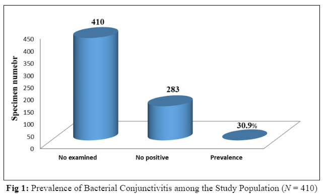
The present study has shown that the percentage prevalence of bacterial conjunctivitis in the study population is 30.9%. Staphylococcus aureus showed the highest prevalencebeing responsible for 47.1% of all cases followed by Escherichia coli responsible for 17.6% of the cases. This is in keeping with some other studies which have demonstrated the same trend, Esenwah, 2005 [11], in a research carried out in Owerri of Nigeria reported that Staphylococcus and E. coli microorganisms are the most prevalent microorganisms responsible for most external eye infections such as conjunctivitis in Owerri. Likewise research carried out in Ibadan of Nigeria reported that the common bacterial pathogen of conjunctivitis in that environment was Staphylococcus aureus, being responsible for 74.9% of all cases [6]. This increase in the prevalence of Staphylococcus aureus can be attributed to the contamination of the eye from skin normal flora as a result of touching the eyes with contaminated hands. It may also be attributed to the fact that Staphylococcus aureus has the ability to multiply and spread more widely in tissues than any other common bacterial cause of conjunctivitis through their production of many extracellular substances like coagulase. The normal microflora of the eye, which resembles that of the skin primarily, consists of staphylococci, coryneform bacteria and some other anaerobes [12]. But Fahmy et al., [13] demonstrated that cultures of the normal eye are frequently sterile though Staphylococcus aureus may be seen occasionally but in very small numbers [13]. Therefore, increased number or significant growth of Staphylococcus may suggest manifest inflammation in the eye [13]. In this study, E. coli on the other hand accounted for 17.6% of the cases. It is also found as part of the normal flora of the intestinal tract of humans and animals, in the soil and as plant pathogens. E. coli had been reported to be responsible for certain ocular inflammations such as conjunctivitis [14]. Thus, conjunctival infection with E. coli can be attributed toits easily spread through scratches with the fingernails and rubbing of the eye with infected hands. This study also showed that males were more infected with bacterial conjunctivitis than females with significant difference (p = 0.003). This is in agreement with some previous reports [15] while some previous workers found a high prevalence of the disease in female patients as Omar et al., 2016 reported that females were more infected than males [16]. This can be explained by gender variation that may vary on a region to region basis [17]. However, the higher percentage prevalence in males can also be attributed to occupation, as males were more involved in outdoor activities which predisposes to ocular infections either directly or following trauma than females such as farming. This study also shows that the highest prevalence was among infants and children of age group less than 15 years old (58.8%). The age frequency was statistically significant (p = 0.009). Okesola and Salako, 2010, in a study carried out in Ibadan reported the highest rate of bacterial conjunctivitis 96(26.3%) was found among infants and children [6]. This is also in agreement with the report by Lichtenstein and Rinehart, 2003 in the United States that bacterial conjunctivitis is more prevalent in children than in adults 28% [18]. This higher percentage prevalence in infants and children can be attributed to the fact that children play more aggressively in dusty contaminated environments and probably due to lack of good hygiene. Oppositely Abdullah et al., found that conjunctivitis was more common among elderly (61 to 70 years) with 34.4% [19]. The present study indicated that the maximum antibiotic sensitivity of all the isolates was against gentamicin (100%) followed by ciprofloxacin 29/34 (85.3%) followed by chloramphenicol 27/34 (79.4%). This is similar to one study that reported all of the bacterial isolates of conjunctivitis were susceptible to chloramphenicol, ciprofloxacin and gentamicin [20]. Similarly, in a study conducted in Ile-Ife, Nigeria, reported susceptibility rate of 78.9% to chloramphenicol and 89.8-97.8% to ciprofloxacin, a quinolone [21]. Gentamicin and chloramphenicol have the least side effects and hence are safe to be prescribed even for children. Chloramphenicol is the drug of choice for treatment of infectious conjunctivitis in many countries [16]. Susceptibilities to the remaining antibiotics used in this study were rather poor, 44.1% to ampicillin, 47.1% to erythromycin and 50% to tetracycline. The present study showed that the maximum resistance was against erythromycin (50%) followed by ampicillin (47.1%), but in a study conducted in Ibadan, Nigeria in 2010, reported susceptibility rate of 57% to erythromycin [6]. The low susceptibility to these agents observed in this study, may be attributed to increased rate of abuse of these antibiotics especially ampicillin in our community. Staphylococcus aureus, the most prevalent pathogen in this study, was most susceptible to gentamicin (100%), and least to tetracycline (43.75%) ,an antibiotic that is commonly used for bacterial conjunctivitis in the study location. [Table1, 2, 3, 4, 5, 6, 7]
Table 1: Prevalence of Various Bacterial Agents of Conjunctivitis (N = 34)
Bacterial pathogen
No (%)
Chi-square value
P-value
Staphylococcus aureus
16(47.1%)
105.378
0.00
Coagulase negative staphylococci
4(11.8%)
Escherichia coli
6(17.6%)
Pseudomonas aeruginosa
4(11.8%)
Proteus mirabilis
4(11.8)
Table 2: Distribution of Patients with Conjunctivitis by Age (N = 110)
Age (years)
No (%)
Chi-square value
P-value
<15
20(58.8%)
11.633
0.009
16-30
8(23.5%)
31-45
2(5.9%)
>45
4(11.8%)
Table 3: Distribution of Patients with Conjunctivitis by Gender (N = 110)
Gender
No (%)
Chi-square value
P-value
Male
22(67.7%)
18.084
0.003
Female
12(35.3%)
Table 4: Antibiotic Susceptibility Pattern of S. Aureus Isolates (N=16)
Antibiotic Name
Sensitive %
Intermediate%
Resistant%
Gentamicin
16(100%)
0(0.0%)
0(0.0%)
Chloramphenicol
12(75%)
1(6.2%)
3(18.8%)
Ampicillin
0(0.0%)
2(12.5%)
14(87.5%)
Erythromicin
1(6.2%)
1(6.2%)
14(87.5%)
Ciprofloxacin
12(75%)
4(25%)
0(0.0%)
Tetracycline
3(18.8%)
4(25%)
9(56.2%)
According to Clinical and Laboratory Standards Institute (CLSI), 2017 [22]
According to Clinical and Laboratory Standards Institute (CLSI), 2017 [22]
Table 6: Antibiotic Susceptibility Pattern of E .Coli Isolates (N=6)
Antibiotic Name
Sensitive %
Intermediate%
Resistant%
Gentamicin
6(100%)
0(0.0%)
0(0.0%)
Chloramphenicol
5(83.3%)
1(16.7%)
0(0.0%)
Ampicillin
6(100%)
0(0.0%)
0(0.0%)
Erythromicin
6(100%)
0(0.0%)
0(0.0%)
Ciprofloxacin
6(100%)
0(0.0%)
0(0.0%)
Tetracycline
6(100%)
0(0.0%)
0(0.0%)
According to Clinical and Laboratory Standards Institute (CLSI), 2017 [22]
Table 7: Antibiotic Susceptibility Pattern of Pseudomonas Aeruginosa Isolates (N=4)
Antibiotic Name
Sensitive %
Intermediate%
Resistant%
Gentamicin
4(100%)
0(0.0%)
0(0.0%)
Chloramphenicol
4(100%)
0(0.0%)
0(0.0%)
Ampicillin
4(100%)
0(0.0%)
0(0.0%)
Erythromicin
4(100%)
0(0.0%)
0(0.0%)
Ciprofloxacin
4(100%)
0(0.0%)
0(0.0%)
Tetracycline
4(100%)
0(0.0%)
0(0.0%)
According to Clinical and Laboratory Standards Institute (CLSI), 2017 [22]
CONCLUSION:
The percentage prevalence of bacterial conjunctivitis in the study population is 30.9%, bacterial agents isolated includes Staphylococcus aureus, coagulase negative Staphylococci, Escherichia coli, Pseudomonas aeruginosa and Proteus mirabilis. The limitation of the current study was that Chlamydia trachomatis, Corynebacterium species and anaerobic bacteria as well as prevalence of methicillin resistance among the isolated S. aureus responsible for bacterial conjunctivitis were not investigated due to resources problems; though, further studies will be conducted in this regard.
Funding: No funding sources
Conflict of interest: None declared
Ethical approval: The study was approved by the Institutional Ethics Committee of Usmanu Danfodiyo University
REFERENCES:
San Francisco, C. A. Francisco. "Cornea/External Disease Panel. Preferred Practice Pattern Guidelines: American Academy of Ophthalmology. Conjunctivitis-Limited Revision: 9-16." (2013).
Sharma, Savithri. "Ocular infections: research in India." Indian journal of medical microbiology 28.2 (2010): 91.DOI:10.4103/0255-0857.62481
Mannis, M. J., and Plotnik, R. D. "Bacterial conjunctivitis." Duane's Ophthalmology on CD-ROM, edited by W. Tasman and E. A. Jaeger, Lippincott Williams & Wilkins, (2006).
Okesola, A. O., and A. O. Salako. "Microbiological profile of bacterial conjunctivitis in Ibadan, Nigeria." Annals of Ibadan postgraduate medicine 8.1 (2010): 20-24.https://www.ajol.info/index.php/aipm/article/view/63953
Kowalski, Regis P., Lisa M. Karenchak, and Eric G. Romanowski. "Infectious disease: changing antibiotic susceptibility." Ophthalmology Clinics of North America 16.1 (2003): 1-9.https://europepmc.org/article/med/12683244
Esenwah, E. "Isolation and Identification of the Microorganisms most Prevalent in External Eye Infections as seen in an Eye Clinic in Owerri." Journal of the Nigerian Optometric Association 12 (2005).https://www.ajol.info/index.php/jnoa/article/view/64449
Fahmy, J. A., S. Møller, and M. Weis Bentzon. "Bacterial flora of the normal conjunctiva I. Topographical distribution." Acta ophthalmologica 52.6 (1974): 786-800.https://doi.org/10.1111/j.1755-3768.1974.tb01115.x
Briuser, J. H., and E. M. Burd. "Principles of Diagnostic Ocular Microbiology. Infections of the Eye." (1996): 69-77.
Khan, M. D., N. Kundi, and N. Saeed. "A Study of 530 cases of vernal conjunctivitis form the North Western Frontier Province of Pakistan." (1986): 111-114.https://www.sid.ir/paper/549376/fa
Brinser, John H., and Eileen M. Burd. "Principles of diagnostic ocular microbiology." Infections of the eye. 2nd edn. Boston: Little, Brown & Co (1996): 69-84.
Lichtenstein, S. J., and M. Rinehart. "Levofloxacin bacterial conjunctivitis study G. efficacy and safety of 0.5% levofloxacin ophthalmic solution for the treatment of bacterial conjunctivitis in pediatric patients." J AAPOS 7.5 (2003): 317-24.
Abdullah, Farhan Essa, Mariya Irfan Khan, and Sadia Waheed. "Current pattern of antibiotic resistance of clinical isolates among conjunctival swabs." Pakistan journal of medical sciences 29.1 (2013): 81.https://www.ncbi.nlm.nih.gov/pmc/articles/PMC3809201/
Sthapit, P. R., et al. "Bacterial conjunctivitis and use of antibiotics in Dhulikhel Hospital-Kathmandu University Hospital." Kathmandu University Medical Journal 9.2 (2011): 69-72.https://www.kumj.com.np/issue/34/67-70.pdf
Clinical and Laboratory Standard Institute (CLSI). Performance Standards for Antimicrobial Susceptibility Testing; Twenty-fourth Informational Supplement (CLSI Document M100-S24). CLSI, 2014. Designs in Medical Research. Indian Journal of Psychological Medicine, vol. 35, no. 2.
Advertisement
Recommended Articles
Research Article
A Critical Analysis of Spinal Block Anesthesia under Ultrasonographic Guided Technique: Is Levobupivacaine Alone Effective or on Adjuvant is Warranted?
Verma AK
Published: 20/09/2021
Download PDF
Cite
x
APA
AK, V. (2021). A Critical Analysis of Spinal Block Anesthesia under Ultrasonographic Guided Technique: Is Levobupivacaine Alone Effective or on Adjuvant is Warranted?. Himalayan Journal of Applied Medical Sciences and Research, 2(2), 1-4.
MLA
AK, Verma. "A Critical Analysis of Spinal Block Anesthesia under Ultrasonographic Guided Technique: Is Levobupivacaine Alone Effective or on Adjuvant is Warranted?." Himalayan Journal of Applied Medical Sciences and Research 2.2 (2021): 1-4.
Chicago
AK, Verma. "A Critical Analysis of Spinal Block Anesthesia under Ultrasonographic Guided Technique: Is Levobupivacaine Alone Effective or on Adjuvant is Warranted?." Himalayan Journal of Applied Medical Sciences and Research 2, no. 2 (2021): 1-4.
Harvard
AK, V. (2021) 'A Critical Analysis of Spinal Block Anesthesia under Ultrasonographic Guided Technique: Is Levobupivacaine Alone Effective or on Adjuvant is Warranted?' Himalayan Journal of Applied Medical Sciences and Research 2(2), pp. 1-4.
Vancouver
AK V. A Critical Analysis of Spinal Block Anesthesia under Ultrasonographic Guided Technique: Is Levobupivacaine Alone Effective or on Adjuvant is Warranted?. Himalayan Journal of Applied Medical Sciences and Research. 2021 Jul;2(2):1-4.
Download PDF
Research Article
The Role of Elevated CRP in Assessing the Severity and as a Prognostic Marker in Covid19 Patients
V. S. Sheshan,
...
Shekhar N. Chandra
Published: 04/07/2021
Download PDF
Cite
x
APA
S. Sheshan, V., Mulla Bahuddeen, M., None, M. & N. Chandra, S. (2021). The Role of Elevated CRP in Assessing the Severity and as a Prognostic Marker in Covid19 Patients. Himalayan Journal of Applied Medical Sciences and Research, 2(2), 1-4.
MLA
S. Sheshan, V., et al. "The Role of Elevated CRP in Assessing the Severity and as a Prognostic Marker in Covid19 Patients." Himalayan Journal of Applied Medical Sciences and Research 2.2 (2021): 1-4.
Chicago
S. Sheshan, V., M. Mulla Bahuddeen, Mahendra and Shekhar N. Chandra. "The Role of Elevated CRP in Assessing the Severity and as a Prognostic Marker in Covid19 Patients." Himalayan Journal of Applied Medical Sciences and Research 2, no. 2 (2021): 1-4.
Harvard
S. Sheshan, V., Mulla Bahuddeen, M., None, M. and N. Chandra, S. (2021) 'The Role of Elevated CRP in Assessing the Severity and as a Prognostic Marker in Covid19 Patients' Himalayan Journal of Applied Medical Sciences and Research 2(2), pp. 1-4.
Vancouver
S. Sheshan V, Mulla Bahuddeen M, Mahendra M, N. Chandra S. The Role of Elevated CRP in Assessing the Severity and as a Prognostic Marker in Covid19 Patients. Himalayan Journal of Applied Medical Sciences and Research. 2021 Jul;2(2):1-4.
Download PDF
Case Report
Case Report: Bilateral Papilledema in Neurocysticercosis
Shaloo Negi,
Bhagwan Dass Negi
Download PDF
Cite
x
APA
Negi, S. & Dass Negi, B. (2021). Case Report: Bilateral Papilledema in Neurocysticercosis. Himalayan Journal of Applied Medical Sciences and Research, 2(1), 1-3.
MLA
Negi, Shaloo and Bhagwan Dass Negi. "Case Report: Bilateral Papilledema in Neurocysticercosis." Himalayan Journal of Applied Medical Sciences and Research 2.1 (2021): 1-3.
Chicago
Negi, Shaloo and Bhagwan Dass Negi. "Case Report: Bilateral Papilledema in Neurocysticercosis." Himalayan Journal of Applied Medical Sciences and Research 2, no. 1 (2021): 1-3.
Harvard
Negi, S. and Dass Negi, B. (2021) 'Case Report: Bilateral Papilledema in Neurocysticercosis' Himalayan Journal of Applied Medical Sciences and Research 2(1), pp. 1-3.
Vancouver
Negi S, Dass Negi B. Case Report: Bilateral Papilledema in Neurocysticercosis. Himalayan Journal of Applied Medical Sciences and Research. 2021 Jan;2(1):1-3.
Download PDF
Research Article
Prevalence of Sacroiliac Joints Involvement in Iraqi Patients with Seropositive Rheumatoid Arthritis
Abbas Hamzah Zainel
Published: 30/12/2024
Download PDF
Cite
x
APA
Hamzah Zainel, A. (2024). Prevalence of Sacroiliac Joints Involvement in Iraqi Patients with Seropositive Rheumatoid Arthritis. Himalayan Journal of Applied Medical Sciences and Research, 5(2), 1-4.
MLA
Hamzah Zainel, Abbas. "Prevalence of Sacroiliac Joints Involvement in Iraqi Patients with Seropositive Rheumatoid Arthritis." Himalayan Journal of Applied Medical Sciences and Research 5.2 (2024): 1-4.
Chicago
Hamzah Zainel, Abbas. "Prevalence of Sacroiliac Joints Involvement in Iraqi Patients with Seropositive Rheumatoid Arthritis." Himalayan Journal of Applied Medical Sciences and Research 5, no. 2 (2024): 1-4.
Harvard
Hamzah Zainel, A. (2024) 'Prevalence of Sacroiliac Joints Involvement in Iraqi Patients with Seropositive Rheumatoid Arthritis' Himalayan Journal of Applied Medical Sciences and Research 5(2), pp. 1-4.
Vancouver
Hamzah Zainel A. Prevalence of Sacroiliac Joints Involvement in Iraqi Patients with Seropositive Rheumatoid Arthritis. Himalayan Journal of Applied Medical Sciences and Research. 2024 Jul;5(2):1-4.
A, N. & A, U. (2021). Bacterial Conjunctivitis among Patients of Different Age Groups Attending Some Eye Clinics in Sokoto, Nigeria. Himalayan Journal of Applied Medical Sciences and Research, 2(2), 1-5.
MLA
A, Nuhu and Usman A. "Bacterial Conjunctivitis among Patients of Different Age Groups Attending Some Eye Clinics in Sokoto, Nigeria." Himalayan Journal of Applied Medical Sciences and Research 2.2 (2021): 1-5.
Chicago
A, Nuhu and Usman A. "Bacterial Conjunctivitis among Patients of Different Age Groups Attending Some Eye Clinics in Sokoto, Nigeria." Himalayan Journal of Applied Medical Sciences and Research 2, no. 2 (2021): 1-5.
Harvard
A, N. and A, U. (2021) 'Bacterial Conjunctivitis among Patients of Different Age Groups Attending Some Eye Clinics in Sokoto, Nigeria' Himalayan Journal of Applied Medical Sciences and Research 2(2), pp. 1-5.
Vancouver
A N, A U. Bacterial Conjunctivitis among Patients of Different Age Groups Attending Some Eye Clinics in Sokoto, Nigeria. Himalayan Journal of Applied Medical Sciences and Research. 2021 Jul;2(2):1-5.