Background: Patients with compound fractures of the lower limb long bones caused by missile injuries are at high risk of Venous Thromboembolism (VTE) due to immobilization, endothelial injury and trauma-induced hypercoagulability. The optimal use of pharmacological thromboprophylaxis in this population remains controversial because of concerns regarding bleeding complications. Objective: To evaluate the effectiveness and safety of pharmacological prophylaxis with low-molecular-weight heparin in preventing Deep Vein Thrombosis (DVT) among patients with compound lower limb long-bone fractures caused by missile injuries, compared with no prophylaxis. Methods: This prospective cohort study was conducted between August 2024 and July 2025 at Al-Kadhimiya Teaching Hospital and Imam Ali Hospital, Baghdad, Iraq. Thirty-three male patients aged 16–48 years with isolated Gustilo type IIIA or IIIB compound fractures of the femur and/or tibia caused by missile injuries were included. Patients were divided into two groups: Group A (n = 15) received subcutaneous enoxaparin (4000 IU once daily) and Group B (n = 18) received no pharmacological prophylaxis. All patients underwent standardized emergency and orthopedic management, including wound debridement and external fixation. Clinical monitoring for DVT and bleeding was performed daily. Doppler ultrasonography was conducted between days 10 and 14 of admission. Data were analyzed using Student’s t-test and Fisher’s exact test, with p <0.05 considered statistically significant. Results: Two patients (11%) in the non-prophylaxis group developed asymptomatic DVT, while no cases were detected in the prophylaxis group. The overall DVT incidence was 6%. The difference between groups was not statistically significant (p = 0.4886). No patients in either group developed clinically significant bleeding or abnormal coagulation profiles. Hemoglobin levels, biochemical parameters and transfusion requirements were comparable between groups. Conclusion: Early administration of enoxaparin appeared to reduce the incidence of DVT in patients with isolated compound lower limb fractures caused by missile injuries without increasing bleeding risk. However, the difference was not statistically significant, likely due to the small sample size. Larger multicenter studies are required to establish definitive evidence and develop standardized thromboprophylaxis protocols for this high-risk trauma population.
Venous Thromboembolism (VTE), which includes Deep Vein Thrombosis (DVT) and Pulmonary Embolism (PE), represents a major public health concern worldwide. The incidence of VTE is approximately 1 per 1,000 person-years and accounts for nearly 5–10% of inpatient hospital deaths in the United States [1]. Notably, VTE-related mortality exceeds that attributed to breast cancer, acquired immunodeficiency syndrome and road traffic accidents combined [1]. Despite being largely preventable, VTE remains one of the most common causes of avoidable morbidity and mortality among hospitalized patients and thromboprophylaxis continues to be underutilized in clinical practice [2]. A large multicenter retrospective study by Schleyer et al. [3] demonstrated suboptimal adherence to VTE prophylaxis guidelines among hospitalized patients. In their analysis of 1,555 medical and surgical intensive care patients across 33 academic medical centers, overall compliance was only 48%, with rates of 59 and 41% among medical and surgical intensive care units, respectively. These findings highlight a substantial gap between evidence-based recommendations and routine clinical implementation. Consequently, VTE prevention has become a national patient safety priority, emphasized by the Surgeon General’s Call to Action [1] and by the Agency for Healthcare Research and Quality, which ranks thromboprophylaxis among the most important patient safety interventions [4]. Nevertheless, limited consensus persists regarding standardized risk assessment models and evidence-based prophylaxis protocols for trauma patients [2]. Orthopedic trauma patients constitute a particularly high-risk population for VTE development. Injury-related factors such as spinal cord injury, pelvic and lower-extremity fractures, high injury severity scores and prolonged mechanical ventilation significantly increase thrombotic risk [5]. Major trauma frequently activates one or more components of Virchow’s triad-hypercoagulability, endothelial injury and venous stasis-thereby creating a highly prothrombotic environment early after injury [6]. This early activation of coagulation pathways complicates both diagnostic evaluation and preventive strategies. The diagnosis of VTE remains challenging because clinical manifestations are often nonspecific and variable. Clinical practice guidelines recommend the use of validated prediction models, such as the Wells score, in combination with D-dimer testing and appropriate imaging modalities [7]. Duplex ultrasonography is the primary diagnostic tool for suspected lower-extremity DVT, while computed tomography pulmonary angiography and ventilation–perfusion scanning are commonly used for suspected PE. However, VTE remains underdiagnosed and a substantial proportion of pulmonary embolism cases are detected only at autopsy. Moreover, routine postoperative duplex ultrasound screening in asymptomatic orthopedic trauma patients has not been shown to reduce PE incidence [8] and is therefore discouraged by major orthopedic societies [9]. The prevention of VTE in trauma patients presents unique clinical challenges. Thrombotic processes often begin immediately following injury, frequently before pharmacologic prophylaxis can be safely initiated. This dilemma is further complicated by concomitant injuries that increase bleeding risk, such as traumatic brain injury and solid organ trauma [2]. A randomized controlled study by Phelan et al. [10] demonstrated that early pharmacologic thromboprophylaxis in patients with traumatic brain injury did not significantly increase the risk of intracranial hemorrhage compared with delayed therapy. Despite their widespread use, mechanical prophylactic methods have shown limited effectiveness in preventing symptomatic DVT and PE in orthopedic patients [11,12]. In the absence of pharmacologic prophylaxis, reported rates of DVT and PE may exceed 50 and 10%, respectively, with fatal pulmonary embolism representing the third leading cause of death among trauma survivors beyond the first day of injury [13]. Pharmacologic prophylaxis, including unfractionated heparin, low-molecular-weight heparins, aspirin and warfarin, has demonstrated substantial efficacy in reducing thromboembolic risk [2]. A systematic review confirmed that pharmacologic thromboprophylaxis significantly reduces the incidence of DVT in hospitalized patients [14]. Current guidelines from the American College of Chest Physicians and the Orthopaedic Trauma Association recommend early initiation of low-molecular-weight heparin in major trauma patients once hemostasis is achieved and bleeding risk is acceptable [9,15]. Recommended durations of therapy vary according to injury type and surgical intervention and individualized clinical judgment remains essential due to limited high-quality evidence. Despite advances in prevention and management, VTE continues to represent a major cause of morbidity and mortality in trauma populations. The absence of universally accepted risk stratification tools and standardized prophylactic protocols underscores the need for further high-quality research. Establishing evidence-based, safe and effective prevention strategies remains essential to improving clinical outcomes in orthopedic and trauma patients [2].
Patients and Methods
This prospective cohort study was conducted from August 2024 to July 2025 at Al-Kadhimiya Teaching Hospital and Imam Ali Hospital, Baghdad, Iraq. The study aimed to evaluate the risk factors and complications associated with femoral shaft fractures in adult patients, with particular emphasis on venous thromboembolism and bleeding outcomes. The study population consisted of young adult patients presenting with compound fractures of the lower limb long bones caused by missile injuries.
Inclusion Criteria
Patients were eligible for inclusion if they met the following criteria:
Acute traumatic injury
Compound fracture(s) of a single lower limb long bone (Gustilo type IIIA or IIIB) caused by missile injuries
Admission and management in the orthopedic ward for at least 10 days
Isolated orthopedic injuries without involvement of other body systems
No documented history of chronic medical comorbidities prior to injury
Exclusion Criteria
Patients were excluded if they met any of the following criteria:
Associated contralateral lower limb injuries and/or spinal injuries
Newly diagnosed comorbidities during hospital admission
Pelvic or spinal fractures
Previous injury to the affected limb or history of deep vein thrombosis
Sampling and Data Collection
The study sample included 33 trauma patients with compound fractures of one lower limb long bone caused by missile injuries, involving the femur, tibia, or both ipsilateral bones. Data were collected by the researcher through direct patient follow-up using a structured data collection form.
The patients’ ages ranged from 16 to 48 years and all participants were male. All patients were initially admitted through the emergency department. Initial evaluation was performed according to the Advanced Trauma Life Support (ATLS) protocol and secondary survey, which confirmed isolated orthopedic injuries.
During emergency management, patients received appropriate antibiotics, analgesics and fluid resuscitation. Laboratory investigations and radiological assessments were performed on admission.
Following emergency stabilization, patients were transferred to the orthopedic ward for further assessment and management. All patients underwent urgent wound debridement and external fixation. Fractures were classified according to the Gustilo classification system and only type IIIA and IIIB injuries were included in the study.
External fixation was applied to the femur, tibia, both bones, or as spanning fixation, depending on the injury pattern. External fixation was used either as temporary stabilization before conversion to internal fixation or as definitive treatment. Management included single or multiple sessions of wound debridement with repeated clinical evaluation.
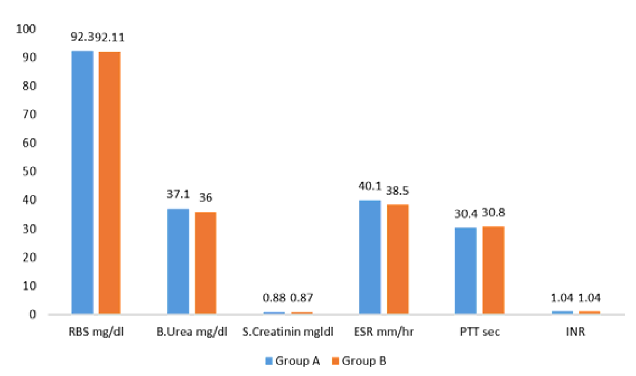
All patients received one or more units of blood transfusion during hospitalization. Emergency laboratory investigations on admission included hemoglobin level, random blood glucose, blood urea and serum creatinine. Follow-up laboratory tests were performed on the second day of admission and during the remaining hospital stay, including complete blood picture, fasting blood glucose, renal function tests and coagulation profile (PT, PTT and INR).
Study Groups and Thromboprophylaxis
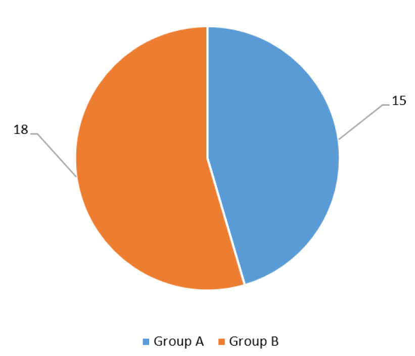
Patients were managed by multiple orthopedic surgeons from different units within the department. According to the treating surgeons’ clinical decisions, patients were divided into two groups:
Patients who received pharmacological thromboprophylaxis
Patients who did not receive pharmacological thromboprophylaxis
Pharmacological prophylaxis consisted of subcutaneous enoxaparin 4000 IU once daily, initiated within 12 hours after urgent surgical intervention. A baseline platelet count was obtained before starting therapy. Additional baseline assessments included patient body weight, coagulation profile, renal function tests and complete blood picture.
Clinical Monitoring and Follow-Up
All patients underwent daily clinical assessment for signs and symptoms of deep vein thrombosis, including:
Leg swelling
Redness
Local warmth
Tenderness
Monitoring for complications of pharmacological prophylaxis was also performed daily, with particular attention to bleeding from the wound site or other anatomical sites.
Between the 10th and 14th day of admission, all patients underwent Doppler ultrasonography of the lower limbs as a screening test for venous thrombosis.
Data were collected throughout the hospitalization period, which ranged from 10 to 14 days.
Outcome Measures
Patients were followed for the following outcome measures:
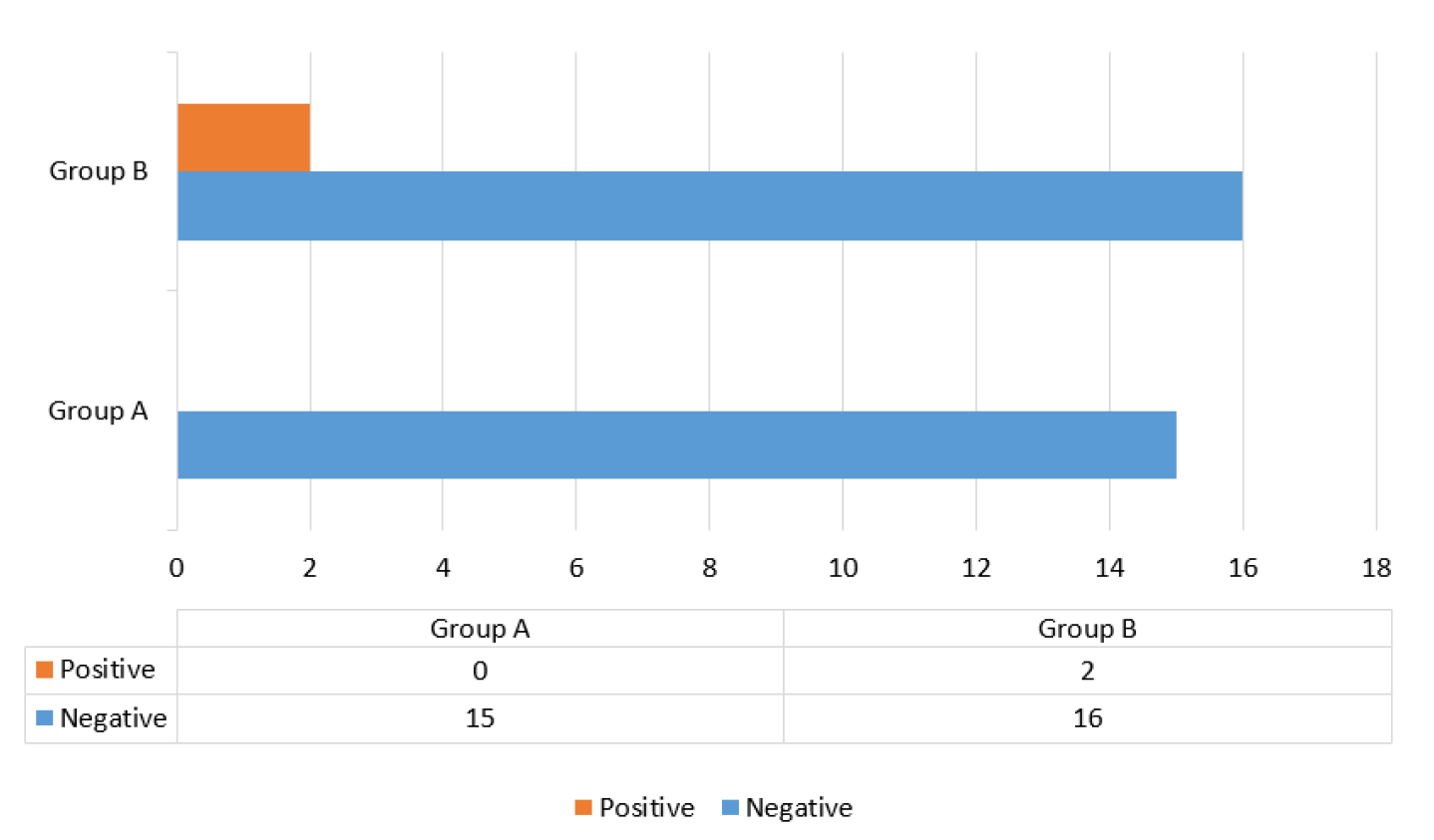
Evidence of deep vein thrombosis, based on clinical signs and symptoms and Doppler ultrasound findings
Evidence of bleeding tendency, defined as bleeding from sites other than the wound and/or abnormal coagulation profile results
Following completion of orthopedic management, patients were either transferred to the plastic surgery unit for further care, discharged home, or referred to nearby hospitals for continued treatment.
Statistical Analysis
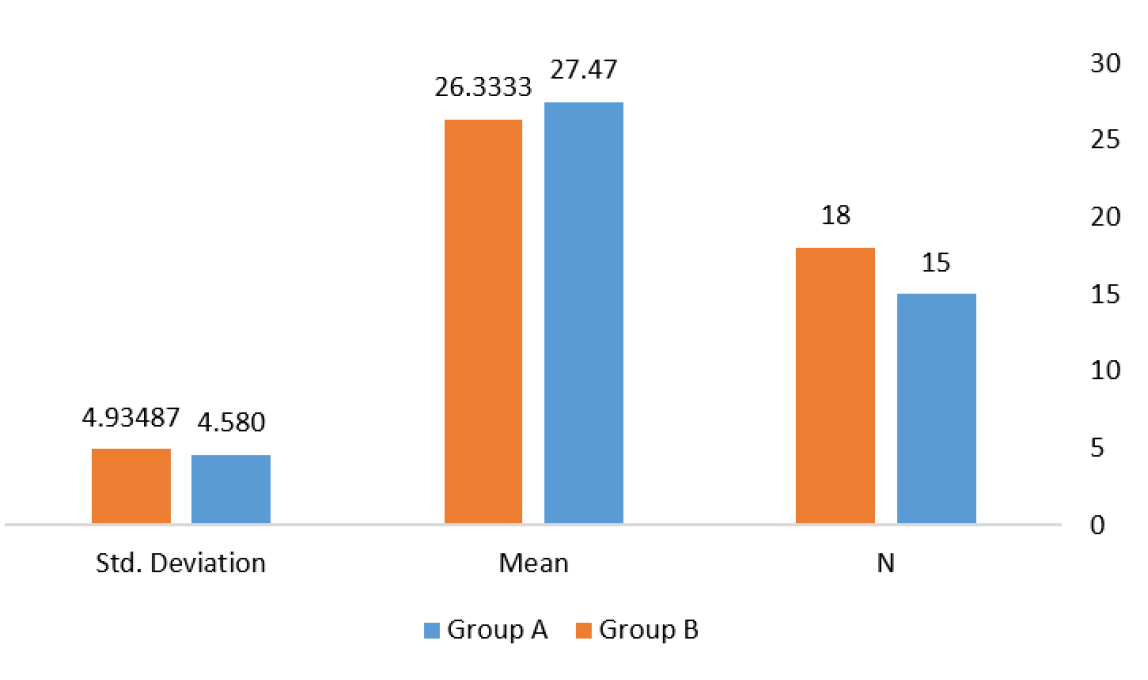
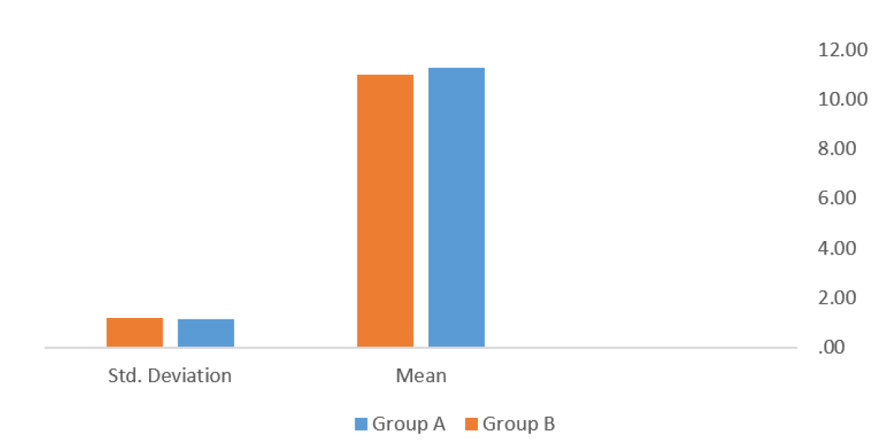
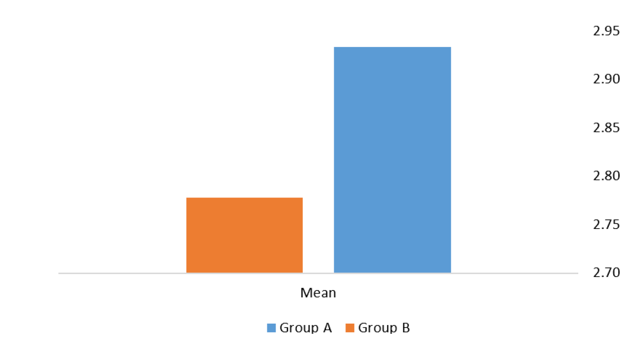
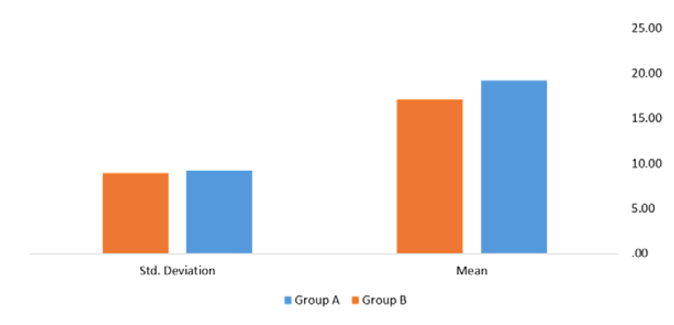
All collected data were entered and analyzed using computerized statistical software. Descriptive statistics were expressed as mean and standard deviation for continuous variables and as frequencies and percentages for categorical variables.
Comparative analyses were performed using Student’s t-test for parametric data and Fisher’s exact test for categorical variables. A p-value of less than 0.05 was considered statistically significant. Results were presented in the form of tables and graphs.
About 33 patients were included in our study, all of them were males with unilateral lower limb compound fractures. The patients were divided into two groups:
Group A: included 15 patients who received pharmacological prophylaxis
Group B: included 18 patients who didn’t receive pharmacological prophylaxis
Figure 1 shows the total number of patients and distribution between study groups.

Figure 1: Total Number of Patients and Distribution
Figure 2 shows the age distribution of both groups, both age group means and standard deviations were identical with no significant statistical difference.

Figure 2: Age Distribution of Both Study Groups
Each patient had three hemoglobin readings, one at admission and another two in the following days of follow up period. The mean of readings was calculated and then the mean of each group was calculated and was shown in Figure 3. There was no significant difference between two groups.

Figure 3: Mean of Hemoglobin Readings in Study Groups
The mean of number of transfused blood pints in each group was calculated and compared in Figure 4. It shows that each patient had received around three blood pints in the follow up period.

Figure 4: Mean of Number of Transfused Blood Pints for each Patient in Both Groups
Each patient had three basic biochemical readings including random blood sugar, blood urea and serum creatinine and bleeding profile. One of them at admission and another two readings in the following days of follow up period. The mean of these readings was calculated and then the mean of each group was calculated and were shown in Figure 5. There was no significant difference between two groups.

Figure 5: The Means of Basic Biochemical Investigations of Both Groups
In Figure 6, the time from injury of each patient to the first operation in hours was calculated and the mean of this time in each group were compared.

Figure 6: Means of Time in Hours from Injury to First Operation
The number of smokers in both groups was shown in Figure 7.

Figure 7: The Number of Smokers in Each Group
The number of positive Doppler findings in the two groups is shown in Figure 8. Two patients in group B had positive Doppler findings of DVT involving the leg and thigh while no occurrence in group A.

Figure 8: The Positive Doppler Findings in Each Group
The incidence of positive Doppler findings in group A was 0% while in group B was 11%. The incidence in both groups is 6%. After application of Fischer exact test for categorical data, the p value equals 0.4886 which isn’t considered to be statistically significant.
In both groups, the patients were clinically asymptomatic regarding DVT with no incidence of bleeding tendency or elevated bleeding profiles.
During the past decade, a substantial increase in trauma incidence has been observed in the study region, with a significant proportion of injuries resulting in compound fractures of the lower extremities. These injuries impose a considerable clinical and socioeconomic burden and require intensive multidisciplinary management. Consequently, improving the quality of care and reducing preventable complications in this patient population have become major priorities. Venous Thromboembolism (VTE) prevention is widely recognized as a critical patient safety issue and comprehensive guidelines exist for elective orthopedic procedures. However, standardized protocols for trauma patients with concurrent bleeding risk remain limited [2]. One of the principal challenges in managing trauma patients lies in balancing physiological hemostasis with the prevention of pathological thrombosis. While activation of the coagulation system is essential for hemorrhage control, excessive activation may predispose patients to venous thromboembolism [16]. Achieving an optimal balance between bleeding and thrombosis continues to be a major research focus (17), providing the rationale for the use of pharmacological thromboprophylaxis in high-risk patients. Lower limb compound fractures fulfill all components of Virchow’s triad, thereby creating a highly thrombogenic environment [6]. Immobilization resulting from fractures and external fixation contributes to venous stasis, while endothelial injury may occur directly from trauma or indirectly through compartment syndrome, infection and repeated surgical interventions. Furthermore, trauma-related hypercoagulability may be exacerbated by multiple blood transfusions, consumption coagulopathy and infection-related prothrombotic states, including methicillin-resistant Staphylococcus aureus–associated thrombosis. These mechanisms collectively place patients with compound fractures at particularly high risk for VTE. Knudson et al. identified immobilization exceeding three days, age ≥30 years and pelvic or lower-extremity fractures as major predictors of venous thromboembolism in trauma patients [18]. In the present study, participants were selected to minimize confounding variables, as all patients had isolated unilateral compound fractures without additional systemic injuries or comorbidities. This design allowed for focused evaluation of fracture-related thrombotic risk and the effect of pharmacological prophylaxis. The study population comprised 33 male patients with unilateral compound fractures of the femur, tibia, or both bones caused by missile injuries. All patients underwent standardized emergency management, including wound debridement and external fixation. However, variability in clinical practice existed among treating surgeons regarding the routine use of anticoagulant prophylaxis. Consequently, patients were divided into two groups: those who received enoxaparin and those who did not. Low-Molecular-Weight Heparin (LMWH) was selected due to its established efficacy, predictable pharmacokinetics and favorable safety profile compared with unfractionated heparin [19]. Mechanical prophylaxis methods, such as intermittent pneumatic compression devices, were not feasible in this cohort due to open wounds, external fixation and soft tissue injuries. Similar limitations have been reported in previous trauma studies, highlighting the practical challenges of implementing mechanical prophylaxis in patients with compound fractures. Daily clinical assessment for DVT was performed in all patients. However, clinical diagnosis alone is known to be unreliable, as many patients with DVT remain asymptomatic [20,21]. Therefore, Doppler ultrasonography was used as the primary screening modality. Since the 1990s, duplex ultrasound has become the preferred noninvasive diagnostic tool for DVT detection [8]. Barrera et al. [14] reported that duplex ultrasound was the most commonly used diagnostic method in randomized trials evaluating thromboprophylaxis. In the present study, Doppler ultrasound was selected due to its safety, sensitivity and availability within the study hospitals. Post-traumatic thrombus formation may occur within 24 hours of injury and may affect both injured and uninjured limbs [21]. In this study, Doppler examinations were performed between 10 and 14 days after injury, reflecting logistical constraints and patient clinical status. Although earlier screening might have detected additional cases, the chosen timing allowed assessment during the peak risk period associated with immobilization and early postoperative recovery. The study identified two cases of asymptomatic DVT in the non-prophylaxis group and none in the enoxaparin group, resulting in an incidence of 11% in untreated patients and 6% overall. These findings are comparable to those reported by Geerts et al. who documented an 18% DVT rate among trauma patients who did not receive prophylaxis [22]. Furthermore, a meta-analysis involving 997 trauma patients demonstrated that prophylaxis significantly reduced DVT risk without increasing bleeding complications [14]. Knudson et al. [23] reported DVT rates of 12% in patients using sequential compression devices and 8% in those receiving low-dose heparin. Although their findings support the protective effect of pharmacologic prophylaxis, differences in study design limit direct comparison. Their study included patients with multiple injuries, broader age ranges, longer hospital stays and coagulation abnormalities, all of which may have increased thrombotic risk. Additionally, unfractionated heparin was used instead of LMWH, which may influence efficacy and safety outcomes. In the present study, statistical analysis using Fisher’s exact test revealed no significant association between enoxaparin administration and DVT incidence (p = 0.48). Consequently, the study failed to reject the null hypothesis. Although fewer DVT cases were observed in the prophylaxis group, the difference did not reach statistical significance.
This finding is most likely attributable to the limited sample size and low event rate. Several factors contributed to the small study population. Many patients were transferred to hospitals in other cities after stabilization, some surgeons declined routine anticoagulant use and Doppler ultrasound was not consistently available. As a result, a considerable number of eligible patients could not be included. These limitations reduced the statistical power of the study and restricted the ability to detect modest treatment effects. Despite the lack of statistical significance, the absence of bleeding complications in the enoxaparin group supports the safety of early pharmacological prophylaxis in carefully selected trauma patients. This observation is consistent with previous reports demonstrating the favorable safety profile of LMWH in trauma settings [10,14]. Overall, the findings suggest that early administration of enoxaparin may reduce the incidence of DVT in patients with isolated compound lower limb fractures, although this effect could not be demonstrated conclusively in the present study. Larger, multicenter studies with extended follow-up periods are required to establish definitive evidence and optimize prophylactic strategies in this high-risk population.
Ethical Approval
Written informed consent was obtained from all patients or their legal guardians. Permission was obtained from the responsible surgeons and hospital administration. Institutional approval was secured prior to study initiation and patient confidentiality was strictly maintained throughout the study.
Office of the Surgeon General (US). The Surgeon General’s Call to Action to Prevent Deep Vein Thrombosis and Pulmonary Embolism. U.S. Department of Health and Human Services, 2008.
Wigner, N.A. and D.J. Donegan. “Current Concepts in Deep Vein Thrombosis and Pulmonary Embolism after Trauma.” Orthopedics, vol. 25, no. 3, 2014.
Schleyer, A.M. et al. “Adherence to Guideline-Directed Venous Thromboembolism Prophylaxis among Medical and Surgical Inpatients at 33 Academic Medical Centers in the United States.” American Journal of Medical Quality, vol. 26, 2011, pp. 174–180.
Sobieraj, D.M. et al. Venous Thromboembolism in Orthopedic Surgery: Comparative Effectiveness Review. Agency for Healthcare Research and Quality, 2012.
Baldwin, K. et al. “Venous Thromboembolism in Patients with Blunt Trauma: Are Comprehensive Guidelines the Answer?” American Journal of Orthopedics, vol. 40, 2011, pp. E83–E87.
Dickson, B.C. “Venous Thrombosis: On the History of Virchow’s Triad.” University of Toronto Medical Journal, vol. 81, 2004, pp. 166–171.
Snow, V. et al. “Management of Venous Thromboembolism: A Clinical Practice Guideline from the American College of Physicians and the American Academy of Family Physicians.” Annals of Internal Medicine, vol. 146, no. 3, 2007, pp. 204–210.
Moed, B.R. et al. “Sequential Duplex Ultrasound Screening for Proximal Deep Venous Thrombosis in Asymptomatic Patients with Acetabular and Pelvic Fractures Treated Operatively.” Journal of Trauma and Acute Care Surgery, vol. 72, 2012, pp. 443–447.
American Academy of Orthopaedic Surgeons. Preventing Venous Thromboembolic Disease in Patients Undergoing Elective Hip and Knee Arthroplasty: Clinical Practice Guideline. AAOS, 2011.
Phelan, H.A. et al. “A Randomized, Double-Blinded, Placebo-Controlled Pilot Trial of Anticoagulation in Low-Risk Traumatic Brain Injury: The DEEP I Study.” Journal of Trauma and Acute Care Surgery, vol. 73, 2012, pp. 1434–1441.
Patel, N. et al. “Antiembolism Stockings: A Review.” Journal of Orthopaedic Surgery, vol. 21, no. 3, 2013, pp. 361–364.
Morris, R.J. and J.P. Woodcock. “Intermittent Pneumatic Compression or Graduated Compression Stockings for Deep Vein Thrombosis Prophylaxis? A Systematic Review.” Annals of Surgery, vol. 251, 2010, pp. 393–396.
Geerts, W.H. et al. “Prevention of Venous Thromboembolism: The Seventh ACCP Conference on Antithrombotic and Thrombolytic Therapy.” Chest, vol. 126, no. 3 suppl., 2004, pp. 338S–400S.
Barrera, L.M. et al. “Thromboprophylaxis for Trauma Patients.” Cochrane Database of Systematic Reviews, no. 3, 2013, article CD008303.
Falck-Ytter, Y. et al. “Prevention of VTE in Orthopedic Surgery Patients: ACCP Evidence-Based Clinical Practice Guidelines (9th Edn.).” Chest, vol. 141, no. 2 suppl., 2012, pp. e278S–325S.
Smith, T.O. et al. “Thromboprophylaxis Following Major Skeletal Trauma: A Systematic Review.” European Journal of Trauma and Emergency Surgery, vol. 37, 2011, pp. 479–490.
Velmahos, G.C. et al. “Prevention of Venous Thromboembolism after Injury: An Evidence-Based Report-Part I.” Journal of Trauma, vol. 49, no. 1, 2000, pp. 132–138.
Knudson, M.M. et al. “Prevention of Venous Thromboembolism in Trauma Patients.” Journal of Trauma, vol. 37, no. 3, 1994, pp. 480–487.
Rogers, F.B. et al. “Practice Management Guidelines for the Prevention of Venous Thromboembolism in Trauma Patients.” Journal of Trauma, vol. 53, 2002, pp. 142–164.
Meissner, M.H. “Diagnosis of Venous Thromboembolic Disease in Cancer Patients.” Seminars in Thrombosis and Hemostasis, vol. 29, no. 1, 2003, pp. 11–18.
Freeark, R.J. et al. “Posttraumatic Venous Thrombosis.” Archives of Surgery, vol. 95, no. 4, 1967, pp. 567–575.
Geerts, W.H. et al. “A Prospective Study of Venous Thromboembolism after Major Trauma.” New England Journal of Medicine, vol. 331, no. 24, 1994, pp. 1601–1606.
Knudson, M.M. et al. “Thromboembolism Following Multiple Trauma.” Journal of Trauma, vol. 32, no. 1, 1992, pp. 2–11.