Neurocysticercosis, the infection caused by the larval form of the tapeworm Taenia solium, is the most common parasitic disease of the central nervous system and the most common cause of acquired epilepsy worldwide. Visual loss in NCC is a serious consequence and can result either from direct ocular involvement or secondary to CNS involvement. Here we report a case of neurocysticercosis with bilateral papilledema.
Neurocysticercosis, the infection caused by the larval form of the tapeworm Taenia solium, is the most common parasitic disease of the central nervous system and the most common cause of acquired epilepsy worldwide. This has primarily been a disease that remains endemic in low-socioeconomic countries [1]. Visual loss in NCC is a serious consequence and can result either from direct ocular involvement or secondary to CNS involvement. It can affect any part of the visual pathway from the eye ball, optic nerve to the visual cortex. Optic nerve affection in NCC can be secondary to compression of the optic nerve in the optic canal and optic chiasma or due to a variable duration of raised intracranial tension. Papilledema, unilateral optic atrophy and bilateral optic atrophy have
28 years old male presented with history of headache and gradual loss of vision in both eyes for 15 days. It was associated with complaints of nausea and vomiting. His headache was intense on awakening and bending down position. There was no history of unconsciousness, seizures, or change in behavior. He was smoker; he was non vegetarian by diet. There was no significant history of similar illness in his family. There was no significant social and environmental history. On examination he was healthy and well oriented to time, place and person. His visual acuity was 6/12 in both eyes. Pupillary reaction was normal in both eyes. Intraocular pressure was 16 mmHg in both eyes. Slit lamp examination showed normal anterior segment. Extraocular movements were normal and colour vision was normal in both eyes. A dilated fundus examination revealed blurred, elevated disc margin, obliterated cup, dilated tortous vessels and peripapillary hemorrages. There was obscuration of all vessels on the disc. Findings were consistent with papilledema (stage v frisen scale classification) Figure 1. Visual field analysis showed an enlarged blind spot in both eyes. His vital signs and systemic examination were normal.
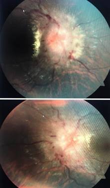
Figure 1: Fundus photograph of right and left eye of patient of neurocysticercosis showing stage v papilledema
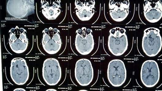
Figure 2: CT scan showing calcified lesion in frontal lobe of brain
Blood reports were hemoglobin 13 gm%, Neutrophils (N) 57%, Lymphocytes (L) 20%, Eosinophils (E) 14%, Monocytes (M) 1%, Erythrocyte Sedimentation Rate (ESR) 30 mm/first hour, Total Leukocyte Count (TLC) 7600 cells/mm3 and Random Blood Sugar (RBS) 110 mg%. His serology was negative. His urine routine microscopy was normal. A CT scan showed calcified lesion in frontal lobe (Figure 2).
A physician consultation was done and he was treated with tablet albendazole 400 bd for 1 month and tablet valproic acid was prescribed 300 mg bd for 1 month. He was kept on follow up.
Taenia solium, the pork tape worm, is a two-host zoonotic cestode. Man harbors the adult tape worm in the small intestine and is the only known definitive host. As in all cestodes, the gravid proglottids at the terminal end of the worm are full of eggs, which are the source of infection with the larval stage, or cysticercosis. The pig is the natural intermediate host, which harbors larval cysts anywhere in its body. Humans become infected with cysts by accidental ingestion of T. solium-infective eggs by feco-oral contamination. In such a case, it preferentially infests the CNS. Symptomatic disease occurs almost exclusively from invasion of the CNS and the eye [3-5]. Seizures and epilepsy are considered to be the most common manifestations of NCC. However, several other neurological disorders can also occur [6]. The symptoms may be a result of obstructive hydrocephalus from blockage occurring in the CSF drainage system. Associated symptoms are those of elevated intracranial pressure such as headache, nausea, vomiting, altered mental status, decreased visual acuity (papilledema). Seizures and focal neurological signs are less frequent in such cases. But, most cases of NCC (70-90%) present with seizures [7]. As of neuroimaging, MRI had been proven to be superior over CT scan in detection and characterization of both parenchymal and ventricular NCC [8].
In the series of 23 cases with visual impairment reported by Chang et al., 13 cases were found to be due to optic neuropathy secondary to papilledema [9]. Proaño et al. [9] in their series of giant neurocyticercosis, encountered a case of bilateral optic atrophy due to nerve entrapment secondary to basal arachnoididtis [9]. Similarly, Pansey et al. reported a 5-year-old child presenting with headache and blindness and bilateral optic atrophy who recovered completely after antiparasitic treatment [10]. Dass et al. reported a child with diminished vision due to bilateral optic atrophy resulting from hydrocephalus caused by NCC [11]. Repeat neuroimaging is warranted every 6 months following initial antiparasitic therapy until resolution of cystic lesion. A persistent viable or enhancing lesions on follow-up imaging warrant a repeat course of antiparasitic therapy. Improvement in neuroimaging was visible within a month of combination therapy (albendazole + symptomatic therapy) as documented in literature [12].
Differential diagnosis of NCC should always be considered when encountering patients presented with papilledema accompanied by solitary cortical brain lesion, particularly of those who came from an endemic area. An appropriate treatment of antihelminthic and corticosteroid are the mainstay of treatment.
Del Brutto et al. Neurocysticercosis: A Clinical Handbook. Swets & Zeitlinger, 1998.
Chang, Gregory Youngnam and James R. Keane. “Visual loss in cysticercosis: Analysis of 23 patients.” Neurology, vol. 57, no. 3, August 2001, pp. 545-548. http://dx.doi.org/10.1212/wnl.57.3.545.
Capello, M. et al. "Principles and Practice of Pediatric Infectious Diseases." Edited by S.A. Long and L.K. Pickering, Churchill Livingstone, 2008, pp. 1321–1327.
García, Hector H., et al. “Current consensus guidelines for treatment of neurocysticercosis.” Clinical Microbiology Reviews, vol. 15, no. 4, October 2002, pp. 747-756. http://dx.doi.org/10.1128/cmr.15.4.747-756.2002.
Singhi, P. and S. Singhi. "Neurocysticercosis in children." Journal of Child Neurology, vol. 19, 2004, pp. 482–492.
Varma, A. and K.J.B.S. Gaur. "The clinical spectrum of neurocysticercosis in the Uttaranchal region." Journal of the Association of Physicians of India, vol. 50, 2002, pp. 1398–1400. https://pubmed.ncbi.nlm.nih.gov/12583469/
Serpa, Jose A., et al. “Neurocysticercosis in Houston, Texas: An Update.” Medicine, vol. 90, no. 1, January 2011, pp. 81-86. http://dx.doi.org/10.1097/md.0b013e318206d13e.
Zhao, Jing Long, et al. “Imaging spectrum of neurocysticercosis.” Radiology of Infectious Diseases, vol. 1, no. 2, March 2015, pp. 94-102. http://dx.doi.org/10.1016/j.jrid.2014.12.001.
Proaño, Jefferson V., et al. “Medical treatment for neurocysticercosis characterized by giant subarachnoid cysts.” New England Journal of Medicine, vol. 345, no. 12, September 2001, pp. 879-885. http://dx.doi.org/10.1056/nejmoa010212
Pansey, B.K., et al. “Medically treated neurocysticercosis in a blind child. A case report.” Surgical Neurology, vol. 32, no. 1, July 1989, pp. 56-58. http://dx.doi.org/10.1016/0090-3019(89)90036-0.
Dass, Rashna, et al. “Neurocysticercosis presenting as hydrocephalus and bilateral optic atrophy.” Journal of Pediatric Neurosciences, vol. 5, no. 1, January 2010, pp. 90-91. http://dx.doi.org/10.4103/1817-1745.66670