Background: The estimated prevalence of HIV related eye diseases in India is reported to be between 8-45%. Dry eye occurs has been seen in later stages of HIV as common anterior segment disorder. Aim: Present study was conducted to prevalence of dry eye in HIV seropositive patients. Material and Methods: The study was a hospital based cross – sectional study where 120 HIV-positive patients registered at the ART centre and referred to Ophthalmology OPD of a tertiary care teaching hospital in Himachal Pradesh, India for ocular complaints were included in the study. Subjects were investigated for dry eye with Rose Bengal staining, Ocular Surface Disease Index score (OSDI), Schirmers test 1 with and without anaesthesia & Tear Film Break Up Time (TBUT). Results: 25 (10.4%) eyes were found positive on rose Bengal staining while OSDI score was found to be positive for dry eye in 67 patients (55.8%). Based on TBUT Mild, moderate and severe dry eye was found in 58 (24.2%), 45(18.8%) and 13 (5.4%). Based on Schirmer-1 test with anaesthesia, dry eye was seen in 81 (33.7%) eyes. Conclusion: Dry eye is very common in HIV patients than general population hence, routine examination of tear function should be an integral part of assessment of HIV and AIDS patients.
Acquired Immunodeficiency syndrome (AIDS) is one of the most feared infectious dis¬ease of the late 20th century. Since its discovery in 1981, AIDS has emerged as a global health problem. Thirty years after its dis¬covery, virus has reached virtually every corner of the globe [1]. India has a large number of patients with AIDS, which is the third largest population of this group in the world [2].
Ocular manifestations of HIV in India were first reported in 1995. Since then, the number of HIV patients has greatly increased. The estimated prevalence of HIV related eye diseases in India is reported to be between 8-45 % [3].
The immune status of the patient is expected to influence the frequency and nature of manifestations in the eye. Partial immune system recovery following initiation of effective antiretroviral therapy may modify clinical presentation. In addition, in one eye, several infections may occur at the same time, rendering diagnosis and therapeutic intervention more difficult [4].
Keratoconjunctivitis sicca or dry eye occurs in later stages of the disease in 10 -20% of the patients [5]. Dry eye by itself may be of a small consideration in view of the other more serious sequelae of HIV infection. However, when considering the importance of tear film in maintaining the ocular surface integrity, it becomes apparent that treatment of dry eye is important to prevent the sight threatening complications such as secondary corneal ulcers or infections [6].
Several studies have been done on the various ocular manifestations of HIV but very few studied has been done to see the incidence of dry eye in HIV patients especially in North-western part of India. Therefore, present study was planned to see the incidence and severity of dry eye in HIV seropositive patients.
Study Design, Settings and Participants
It was a hospital based cross-sectional study conducted over a period of one year from June 2018 to May 2019 in ophthalmology department of a tertiary care teaching hospital in Himachal Pradesh, India. All HIV-positive patients between 18 to 50 year of age group, registered at the ART centre and referred to Ophthalmology OPD for ocular complaints and asymptomatic patients from ART centre were selected randomly for the study. Patient with confounding factors and co morbidities like diabetes, corneal scars, lid abnormalities, contact wearers, SJS, arthritis, any other connective tissue disorder; patients who were already on treatment for DES, oral contraceptive pills, pregnant females and those taking any medications like antidepressants, antihistaminics, anxiolytics which can independently cause dry eye and seriously ill patients who were not able to cooperate for ophthalmological examination was excluded from the study. Total 346 HIV patients were examined but after considering inclusion and exclusion criteria 120 participants were finally included in our study. Every patient was on Highly Active Antiretroviral Therapy (HAART).
Data Collection
After taking written informed consent from the study subjects, data was collected in a predesigned, pre-tested, semi-structured interview schedule in which socio-demographic profile like age, gender, occupation, duration of ART, duration of disease etc were collected. After taking history as per proforma, each patient has undergone detailed ophthalmological examination in the following sequence.
Dry Eye Questionnaire - Ocular Surface Disease Index Score (OSDI)
OSDI questionnaire was administered to all patients and by a single examiner. To those who were non conversant in English, the questions were explained to the patients in their local language. The OSDI questionnaire has 12 items, with each question given a score ranging from 0 (none of the time) to 4 (all of the time). The final score was calculated by multiplying the sum of all the scores by 25 and then dividing the total by the number of questions answered. Scores range from 0 to 100 with 0–12 representing normal, 13–22 representing mild DED, 23–32 representing moderate DED, and ≥33 representing severe DED [7-8].
TBUT (Tear Film Break Up Time)
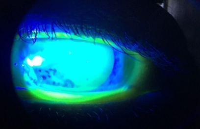
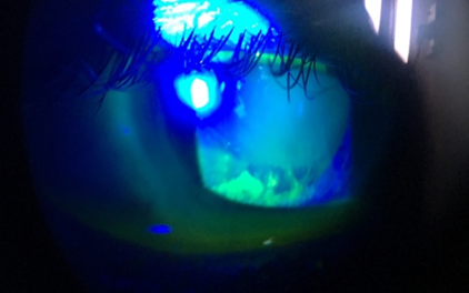
Tear break-up time was performed by moistening a fluorescein strip with sterile non preserved saline and applying it to the inferior tarsal conjunctiva. After several blinks, the tear film was examined using a broad beam of the slit-lamp biomicroscope with a cobalt blue filter. The time lapse between the last blink and the appearance of the first randomly distributed dark discontinuity in the fluorescein -stained tear film is the tear break-up time. Break-up times less than 20 seconds was considered abnormal. This procedure was repeated three times on both eyes [7-9].
Schirmer-1 Test Without Anesthesia
The Schirmer test was performed by placing a narrow filter-paper strip in the inferior cul-de-sac. Aqueous tear production was measured by the length in millimeters that the strip wets during the test period, generally 5 minutes. Test was performed with eyes closed. Serin et al., suggested that administering the Schirmer test with the patient’s eyes closed producesfewer variable results and greater repeatability [10]. While an isolated abnormal result can be nonspecific, serially consistent low results are highly suggestive of aqueous deficiency [8,11-12]. Severity grading was done as follows; >15 mm/5 min (Normal), 10-15 mm/5min (Mild Dry Eye), 5-9 mm/5min (Moderate Dry Eye) and <5 mm/5min (Severe Dry Eye).
Schirmers Test 1 With Anesthesia
The Schirmer test with anesthesia, also referred to as a basic secretion test, has been reported to give more variable results than the Schirmer test done without anesthesia. If topical anesthesia is applied, excess fluid should be gently removed from the cul-de-sac prior to insertion of the filter paper. This test is performed after 15 minutes of Schirmer’s test 1. After putting topical proparacaine drops, schirmer’s strip was applied as for Schirmer’s test 1. Results noted down after 5 minutes. <6mm/5min was considered abnormal and >6 mm of wetting after 5 minutes was considered normal.
Rose Bengal Staining
Rose Bengal staining was done and graded as van bitzerveld scoring. Rose Bengal score > 4 was considered abnormal and < 4 was considered normal. Maximum score was 9. Intensity scored in 2 exposed nasal conjunctival zones and cornea. It was started after putting anaesthetic drop to avoid irritation as this stain is slightly toxic to epithelial cells [7,8].
Posterior segment examination was also performed in every patient. Those patients who were found to have abnormalities other than dry eye were treated accordingly.
Statistical Analysis
Data were analyzed and statistically evaluated using SPSS software, version 17 (Chicago II, USA). Quantitative data was expressed in mean, standard deviation or median with interquartile range while qualitative data were expressed in percentage.
Ethical Issues
All participants were explained about the purpose of the study. Confidentiality was assured to them along with informed written consent. The study was approved by the Institutional Ethical Committee
Out of 120 patients, 67 were males (55.8%) and 33 were females (44.2%). Mean Schirmer-1 test without anaesthesia score was 16.88±5.20 mm, Schirmer-1 test with anaesthesia score was 8.30±2.29 mm, mean TBUT time 16.69±6.79 seconds and mean OSDI score was 17.88±12.09. In 40 (33.3%) patients, CD4 count was > 500 cells/mm3 were, in 50 (41.7%) patients it was between 200 to 500 cells/mm3 and in 30 (25%) patients, CD4 cell count was <200 cells/mm3. Median CD4 count of the patients was 419 (range 21-1387) cells/mm3. On rose Bengal staining, 25 (10.4%) eyes were found positive while OSDI score was found to be positive for dry eye in 67 patients (55.8%) amongst which mild, moderate and severe dry eye was seen in 39 (32.5%), 17 (14.2%) and 11 (9.2%) patients respectively. TBUT (Tear Film Breakup Time) was performed after dry eye questionnaire. It was normal in 124 patients (51.7%) while Mild, moderate and severe dry eye was found in 58 (24.2%), 45(18.8%) and 13 (5.4%) number of patients based on TBUT. Based on Schirmer-1 test without anesthesia, 165 (68.8%) eyes of patients were found normal while 31.2% (75) eyes were found to be abnormal from this test out of which 45 (18.8%), 23(9.6%) and 7(2.9%) had mild, moderate and severe dry eye respectively. Based on Schirmer-1 test with anaesthesia, dry eye was seen in 81 (33.7%) eyes (Table 1).
Table 1: Dry Eye Based on Different Tests in HIV Patients
Tests | No. | % |
Ocular Surface Disease Index (OSDI) score (n=120) | ||
0-12 (Normal) | 53 | 44.2 |
13-22 (Mild dry eye) | 39 | 32.5 |
23-32 (Moderate dry eye) | 17 | 14.2 |
>33 (severe dry eye) | 11 | 9.2 |
Rose Bengal staining (n=240) | ||
Positive | 25 | 10.4 |
Negative | 215 | 89.6 |
Tear Film Breakup Time (TBUT) (n=240) | ||
>20 sec (Normal) | 124 | 51.7 |
10-20 sec (Mild dry eye) | 58 | 24.2 |
5-9 sec (Moderate dry eye) | 45 | 18.8 |
<5 sec (Severe dry eye) | 13 | 5.4 |
Schirmer-1 test without anesthesia (n=240) | ||
>15mm/5 min (Normal) | 165 | 68.8 |
10-15 mm/5 min (Mild dry eye) | 45 | 18.8 |
5-9 mm/5 min (Moderate dry eye) | 23 | 9.6 |
<5 mm/5 min (severe dry eye) | 7 | 2.9 |
Schirmer-1 test with Anesthesia (n=240) | ||
>6mm/5 min (Normal) | 159 | 66.3 |
<6 mm/5 min (abnormal) | 81 | 33.7 |
Figure 1: Photographs 1 And 2 Showing TBUT of the Patient. Photograph 1 Showing TBUT< 5sec in Right Eye And 12 Sec in Left Eye in Photograph 2
Ocular involvement in AIDS is very common and includes various clinical presentations [13]. In addition to the posterior segment lesions, the anterior segment and ocular surface lesions can be vision threatening also. More than 50% of HIV infected patients have anterior segment manifestations. Kerato-conjunctivitis sicca has been reported as one of the most common anterior segment manifestations. Various studies conducted on normal population have shown that the prevalence of dry eye ranges between 10% and 20% [5,14-16].
The etiology of dry eye disease in HIV patients is usually thought to be due to HIV-mediated lymphocytic infiltration of the lacrimal gland. This leads to the destruction of lacrimal acini and the ductal system, as well as direct conjunctival damage [17]. Dry eye can also be due to blepahritis in these patients, pathogenesis of which explained as reduced ability in these patients to control normal flora and more complex changes in cutaneous glands of eyelids. Meibomitis has been the causative factor in lipid layer dysfunction in these patients.[18] The resulting keratoconjunctivitis contributes to a chronic inflammatory state, further promoting cytokine secretion, destruction and dysfunction of the lacrimal gland, and loss of tear production [19].
Other mechanism for DED causation in PLWHA is loss of neural support that promotes glandular atrophy, and cellular breakdown proteins go to the cell surface and further activate T-cells. Increased expression of intercellular adhesion molecule 1 (ICAM-1) and lymphocyte function-associated antigen 1 (LFA-1) in keratoconjunctivitis, causing antigen presentation of T-cells [20].
In our study, 25 (10.4%) eyes were found positive on rose Bengal staining while OSDI score was found to be positive for dry eye in 67 patients (55.8%). Based on TBUT Mild, moderate and severe dry eye was found in 58 (24.2%), 45(18.8%) and 13 (5.4%). Based on Schirmer-1 test without anaesthesia, 165 (68.8%) eyes of patients were found normal while 31.2% (75) eyes were found to be abnormal from this test out of which 45 (18.8%), 23(9.6%) and 7(2.9%) had mild, moderate and severe dry eye respectively. Based on Schirmer-1 test with anesthesia, dry eye was seen in 81 (33.7%) eyes. Finding of our study were in concordance with different studies.
Lucca JA et al. [21] reported 21.4% prevalence of dry eye in HIV and AIDS patients. However, their study was based on individual symptoms of dry eye and the clinical examination used a non-standardized test. De Carlo DK et al. [22] also reported prevalence of dry eye as 38% in HIV infective patients which matched with our results with TBUT. Another study by Geier SA et al. [23] data demonstrated that decreased tear production occurred in approximately 20% – 25% of patients with HIV infection, but decrease in tear production was not found to be associated with the severity of HIV disease. However, in our study it was found that decrease tear production occurred to the extent of up to 56% as from OSDI and 30 % from Schirmer-1 test which is also in similar to this study [23].
In contrast to our study, Burtin T et al. [24] reported that 70% – 80% of HIV-positive patients presented dry eye symptoms and signs which is very high then our study. A recent study by Gowda et al., showed decreased tear production in 50% of the HIV patients by using Schirmer test. However, in Mathebula SD et al. [25] study results from Schirmer test and TBUT showed prevalence of dry eye in around 80% of HIV patients which was also higher compare to our study.
Finding of this study concluded that dry eye is very common in HIV patients than general population hence, routine examination of tear function should be an integral part of assessment of HIV and AIDS patients.
Acknowledgement
We are grateful to all the participants of the study.
Acharya PK et al. "Ocular manifestations in patients with HIV infection/aids who were referred from the art centre, Hassan, Karnataka, India." Journal of clinical and diagnostic research: JCDR, vol. 6, no. 10, 2012, pp. 1756.
World Health Organization. "AIDS epidemic update." Geneva: WHO, 2017-2018.
Ushalatha B et al. "Ocular manifestations in HIV/AIDS." IOSR-Journal of dental and medical sciences, vol. 15, no. 4, 2016, pp. 27-35.
Aastha S et al. "Ophthalmic manifestations in HIV positive patients and the Indian perspective." Journal of AIDS and clinical research, vol. 6, no. 10, 2015.
Gowda HK et al. "Correlation of CD4 count and severity of dry eye disease in human immunodeficiency virus positive patients." International journal of scientific study, vol. 3, 2015, pp. 68-71.
Upadhyay AK et al. "Ocular lesions associated with human immunodeficiency virus infection." Medical journal armed forces India, vol. 66, no. 3, 2010, pp. 235-8.
TFOS DEWS II. "Definition and classification report." Acta ophthalmologica, vol. 96, no. S261, 2018, pp. 136-40.
"Introduction to the report of the international dry eye workshop (2007)." The ocular surface, vol. 5, no. 2, 2007, pp. 69-70.
Akobiec FA et al. Albert & Jakobiecs: Principles & practice of ophthalmology. W B Saunders Company, 2008.
Serin D et al. "A simple approach to the repeatability of the Schirmer test without anesthesia: eyes open or closed?" Cornea, vol. 26, no. 8, 2007, pp. 903-6.
Krachmer J et al. Cornea. Elsevier, 2017.
Lemp MA et al. "The definition and classification of dry eye disease." Ocular surface, vol. 5, no. 2, 2007, pp. 75-92.
Venkat CM et al. "Ocular manifestation in HIV: a clinical study at tertiary centre." Journal of evolution of medical and dental sciences, vol. 7, no. 11, 2018, pp. 1418-22.
Sharma M et al. "Ocular manifestations in patients attending antiretroviral therapy centre at a tertiary care hospital in Himachal Pradesh, India." The Indian journal of medical research, vol. 147, no. 5, 2018, pp. 496.
Hashemi H et al. "Prevalence of dry eye syndrome in an adult population." Clinical & experimental ophthalmology, vol. 42, no. 3, 2014, pp. 242-8.
Sahai A et al. "Dry eye: prevalence and attributable risk factors in a hospital-based population." Indian journal of ophthalmology, vol. 53, no. 2, 2005, pp. 87-91.
Stewart MW. "Human immunodeficiency virus and its effects on the visual system." Infectious disease reports, vol. 4, no. 1, 2012.
Jeng BH et al. "Anterior segment and external ocular disorders associated with human immunodeficiency virus disease." Survey of ophthalmology, vol. 52, no. 4, 2007, pp. 329-68.
Biswas J et al. "Ocular lesions in AIDS: a report of first two cases in India." Indian journal of ophthalmology, vol. 43, no. 2, 1995, pp. 69.
Banker AS. "Posterior segment manifestations of human immunodeficiency virus/acquired immune deficiency syndrome." Indian journal of ophthalmology, vol. 56, no. 5, 2008, pp. 377.
Lucca JA et al. "Keratoconjunctivitis sicca in female patients infected with human immunodeficiency virus." The CLAO journal, vol. 20, no. 1, 1994, pp. 49-51.
DeCarlo DK et al. "Dry eye among males infected with the human immunodeficiency virus." Journal of the American optometric association, vol. 66, no. 9, 1995, pp. 533-8.
Geier SA et al. "Sicca syndrome in patients infected with the human immunodeficiency virus." Ophthalmology, vol. 102, no. 9, 1995, pp. 1319-24.
Burtin T et al. "Abnormalities of the ocular surface in patients with AIDS." Journal francais d'ophtalmologie, vol. 21, no. 9, 1998, pp. 637-42.
Mathebula SD et al. "Ocular surface disorder among HIV and AIDS patients using antiretroviral drugs." African vision and eye health, vol. 78, no. 1, 2019, pp. 1-7.