Behçet's disease is an uncommon multisystem inflammatory condition characterized by immune-mediated vasculitis affecting vessels of different calibers. Clinically, it presents with recurrent oral and genital ulcerations, dermatological manifestations, and ocular involvement, reflecting its systemic nature. The present study was designed to characterize gut microbial patterns in patients diagnosed with Behçet’s disease and to assess selected immune biomarkers — IL-17A, IL-23, CD4, and CD11b — as potential diagnostic and prognostic indicators. Participants were recruited from Al-Sadr Medical City between December 2024 and September 2025, with ages ranging from 11 to 51 years. Venous blood samples were obtained from patients, and serum concentrations of IL-17A, IL-23, CD4, and CD11b were quantified using the ELISA technique. The study population showed a predominance of male patients, with the highest frequency observed in the 22–31-year age group. Statistical analysis demonstrated significantly elevated serum levels of IL-17A and IL-23 in Behçet’s patients compared with healthy controls. Similarly, CD4 and CD11b expression levels were markedly increased. A strong positive correlation was identified among these immune markers, suggesting coordinated immunological activation. In conclusion, IL-17A, IL-23, CD4, and CD11b may serve as valuable diagnostic and predictive biomarkers in Behçet’s disease, and their interrelationship highlights their potential role in disease pathogenesis and progression.
Behçet’s disease (BD) is a form of vasculitis characterized by periodic inflammatory episodes that result in a variety of distinctive clinical manifestations. The condition most commonly affects the eyes, urogenital mucosa, and skin.The precise etiology remains to be elucidated but some have theorised that a genetically prone or susceptible population, when exposed to environmental triggers, leads to the dysregulation of both auto inflammatory as well as autoimmune processes [1].
Epidemiological studies show significant geographic variations in the prevalence of Behçet's disease, with rates ranging per 100,000 people from 20-420 in Turkey, 1.5-15.9 in Southern Europe, and 0.3-4.9 in Northern Europe [2]. Interestingly, ethnic disparities still persist among migrants with higher prevalence rates or their descendants living in areas with lower prevalence rates [3]. What confirms the role of both genetic and environmental factors. The mortality rate in Behçet's disease increases due to complications of the pulmonary artery, large vessels, nervous system, and digestive system. Uveitis associated with Behçet's disease can cause blindness. Therefore, a better understanding of the causes of Behçet's disease and its pathophysiological mechanisms.
The disease phenotype is motivated by the interaction between genetic susceptibility and both innate and adaptive immunity. Neutrophils are highly active, exhibiting increased chemotaxis and oxygen radical production. Monocytes and macrophages also show increased activation and differentiation, leading to heightened inflammation. Th1 and Th17 cell responses are enhanced, producing cytokines that drive inflammation. Gamma delta T cells are also involved, producing interferon-gamma and interleukin-17. The IL-23/IL-17 axis is a crucial inflammatory pathway, where macrophages and dendritic cells (often CD11b+) produce IL-23, which stimulates CD4+ T cells to differentiate into Th17 cells that secrete IL-17A, causing inflammation in autoimmune diseases. CD11b+ cells can also enhance this pathway [4,5,6].
The presence of microbiota plays an interactive role with autoimmune diseases [7,8], indicating an imbalance in the gut microbiota in patients with Behçet's disease. This imbalance is characterized by a decrease in butyrate-producing bacteria, which may affect T-cell differentiation and epigenetic regulation of immune-related genes, a change in tryptophan-metabolizing bacteria that may be linked to interleukin-22 secretion disorder, and a decrease in bacteria known for their anti-inflammatory properties [9]. Regarding oral microbes, the potential role of Streptococcus bacteria may be through molecular mimicry and neutrophil enzymatic activation. Clinical studies in Behçet's disease have shown that (a) the need for dental care is associated with a more severe course of the disease, and (b) antibiotic-enriched mouthwash alleviates pain and ulcers. Fecal transplantation from Behçet's patients into mouse models led to reduced production of short-chain fatty acids, activation of neutrophils, and Th1/Th17 helper T-cell responses. Recipient mice exhibited exacerbation of experimental autoimmune uveitis, encephalitis, and myelitis [10].
Microbes, particularly gut microbes, are considered a major factor in autoimmune and autoinflammatory diseases, raising questions about their potential role in Behçet's disease. An imbalance in saliva or gut microbes may trigger inflammation by affecting immune responses. Accordingly, this study aims to describe the microbiota of patients with Behçet's disease, evaluation some immunological biomarker example IL-17A, IL-23, CD4 and CD11b as diagnosis and Predictive markers.
Patients
The study included patient from al-sader medical city , Age of persons (11-51years) and collection specimen from December 2024 to September 2025.
Inclusion Criteria (ICBD)
A patient is typically included as having a definite diagnosis of BD if they meet the ICBD criteria, which assigns scores to clinical manifestations. A score of 4 or higher is sufficient for diagnosis. this include Oral Aphthous Ulcers, Genital Aphthous Ulcers, Skin Manifestations, Vascular Manifestations , Central Nervous System (CNS). Positive Pathergy Test (an exaggerated skin inflammatory response 24-48 hours after a needle prick. Diagnosis bank on physician-documented clinical manifestations consistent with Behçet’s disease.
Exclusion Criteria
Presence of an inflammatory bowel diseases, sexual infections, rheumatic disorders, malignancy, hepatitis, liver disease, cancer, autoimmune disease. demographic factors depending on the study, such as being outside a certain age range (e.g., younger than 11 or older than 51), pregnancy, Cystinuria.
Healthy Control
A person who does not have the specific disease or disorder control same patients character in age, sex and lifestyle.
Collection Specimen, Isolation and Identification of Bacteria
Specimen collected from mouth sores, inflammation eyes,skin sores, Genital sores and fecal sample , this specimen cultured on Blood agar, manitol salt agar and Macconkey agar . MacFaddin,(11) approved cultured and microscopic characteristics and biochemical assays were used to identify bacterial isolates. Using the Vitek 2 compact system. The diagnostic was likewise done in line with the manufacturer’s recommendations.
Estimation of Immuno-markers by ELISA Assay
Blood samples from patients with Behçet’s disease were sort out to obtain serum, in which the following cytokines were quantified : IL- 17A and IL-23 (Abcam,USA). Also level measurement CD4 and CD11 b (Bioassay/ china ) . These Immuno-markers measured by ELISA .
Statistical Analysis
Data were analyzed using Student’s t-test, with results accessible as mean values ± SD. Statistical examination was performed using analysis of variance (ANOVA). Pearson’s correlation coefficient was used to evaluate relationships between parameters level in this study. Receiver Operating Characteristic (ROC) curve analysis was employed to determine the sensitivity and specificity of each biomarker. A p-value <0.05 was considered statistically significant.
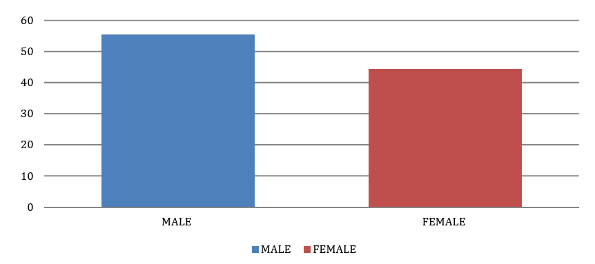
Figure 1 demonstrates the sex proportion of the patients, comprising 15 (55.55%) males and 12 (44.45%) females, with a significant difference (p = 0.001).

Fig 1: Sex Distribution of Behcet Disease Patients
The patients age stretched from 22 to 31 years with higher individuals of age groups; 32-41 years old, the numbers of patients in these age groups were 8( 429.62%) and 7(25.92%) sequentially at P.value 0.0001 . This show in Table 1.
Table 1: Age composition of Behcet Disease participants
p-value | % of BD subjects | NO. of BD subjects | Age |
0.001** | 22.22% | 6 | 11-21 |
29.62% | 8 | 22-31 | |
25.92% | 7 | 32-41 | |
22.22% | 6 | 42-51 | |
100% | 27 | Total |
**Significant correlation at the level of significancy (0.01)
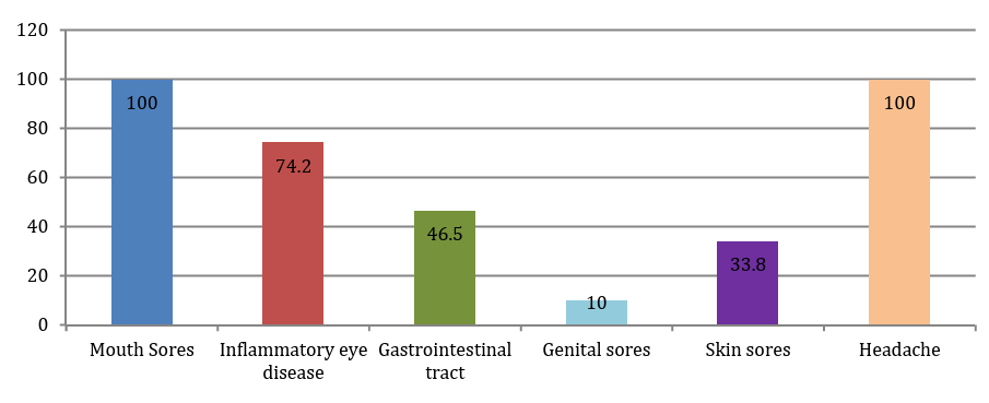
The results of Figure 2 exhibited that the highest Clinical appearances of Mouth Sores has reached100% , while Inflammatory eye disease have look like in second place which 74.2% , while Genital sores have given a 10% incidence , at p-value 0.001.

Figure 2: Clinical Characteristics of Behcets Disease Patients
Among patients specimens, 17 culture-positive samples, this isolates were identified as pathogenic bacteria, defined by exceeding a concentration threshold of 103–104 CFU/mL (14). Cultural morphology, haemolysis, growth on selective media, biochemical test and vitec results appear 93-100%percentage diagnosis bacterial isolates . this bared in Table 2.
Table2: Bacterial Isolates of Behcet Disease Individuals
Bacterial isolates | NO. and percentage | p-value |
Rothia mucilaginosa | 1(5.88%) | 0.001** |
Streptococcus sanguinis | 1(5.88%) | |
Staphylococcus aureus | 3(17.65%) | |
Streptococcus salivarius | 1(5.88%) | |
E.coli | 2(11.76%) | |
lactobacillus | 2(11.76%) | |
Staphylococcus epidermidis | 2(11.76%) | |
streptococcus pyogenes | 3(17.65%) | |
Acinetobacter calcoceticus | 2(11.76%) | |
Total | 17(100%) |
|
Table 3 exposed Information of measurement of some serum Immuno-markers of patients with Behcet disease. The serum cytokine levels for IL-17A and IL-23 had been much higher of patients with Behcet disease (44.375 ±0.021; 7.325±0.12) respectively comparability with controls. CD4 and CD11b have been elevated in Behcet disease (11.19±0.07; 9.97±0.11)respectively compared to controls displayed a significant increase (p<0.05).
Table 3: Immuno-Markers Levels in Participants Compare with Healthy Human
| Cytokines | BD group (N=7) Mean± SE | Healthy group Mean±SE | p-value |
IL-17A | 44.375 ±0.021 | 10.1±0.017 | 0.001** |
IL-23 | 7.325±0.12 | 2.7±0.09 | 0.01* |
CD4 | 11.19±0.07 | 2.15 ±0.08 | 0.02* |
CD11b | 9.97±0.11 | 3.82±0.01 | 0.03* |
In behcet disease patients group, there was very strong positive correlation between different immune-markers (IL-17A, IL-23, CD4 and CD11b ) this show in Table 4.
Table 4: The Correlation Coefficient among IL-17A, IL-23, CD4 and CD11b at Behcet Disease Subjects
Correlation parameters | IL-17A | IL-23 | CD4 | CD11b | |
IL-17A | r | 1 | 0.913** | 0. 846* | 0.932* |
p | - | 0.001 | 0.012 | 0.0001 | |
IL-23 | r | 0.913** | 1 | 0.756** | 0.862* |
p | 0.001 | - | 0.001 | 0.024 | |
CD4 | r | 0. 846* | 0.756** | 1 | 0.972** |
p | 0.012 | 0.001 | - | 0.0001 | |
CD11b | r | 0.932** | 0.862* | 0.972** | 1 |
p | 0.0001 | 0.024 | 0.0001 | - | |
*Significant correlation at the level of significancy (0.05), ** Significant correlation at the level of significancy (0.01)
This study aims to evaluate the potential benefit of some biomarkers as diagnostic and predictive criteria for Behçet's disease, with a particular focus on determining the value of sensitivity and specificity for accurate diagnosis and prediction of the disease. As shown in the Table 5, the level of interleukin-17A was within the optimal range (AUC=0.965) with high a sensitivity 94% and specificity of 90%.
Table 5: ROC analysis of the studied biomarkers in Behçet's disease
Parameters | Sensitivity | Specificity | AUC | 95% Confidence Interval | p-value |
IL-17A | 94% | 90% | 0.965 | 0.935 -0.995 | 0.001** |
IL-23 | 81% | 80% | 0.839 | 0.769- 0.921 | ≤0.001** |
CD4 | 90% | 90% | 0.966 | 0.938- 0.998 | 0.0001** |
CD11b | 100% | 93% | 0.97 | 0.959-1.000 | 0.0001** |
Area Under the Curve (AUC)
On the other hand, the results show that IL-23 also has an optimal range (AUC=0.839) with a sensitivity of (81%) and specificity of (80%). While AUC of CD4 is 0.966 with a sensitivity and specificity which 90%. In addition, CD11b demonstrates a significant perfect area under the curve (AUC= 0.97) with a sensitivity (100%) and specificity of (93%).
The number of Behçet's disease patients is higher among males than females in endemic regions (such as the Middle East and East Asia), and the disease tends to be more severe in men. The specific patterns of organ involvement and disease severity in each sex suggest that a combination of genetic, hormonal, and environmental factors may influence how the disease manifests [12].
Behçet's disease is more common in young adulthood, especially among individuals in their twenties and thirties. Some studies indicate that the most common age for disease onset ranges from 20 to 40 years, with a notable concentration in the 26 to 35 age group in some communities. However, Behçet's disease can occur at any age, although it is rare in young children (childhood onset) or adults over fifty. The disease tends to be more severe in younger patients, particularly young males. Early-onset disease is considered a risk factor for increased disease activity [13].
Patients with Behcet's disease experienced an increase in the frequency of mouth ulcers, as all patients had mouth-and-mouth ulcers. In fact, in most cases, mouth-and-mouth ulcers have been the primary diagnosis for patients. Similarly, mouth-and-mouth ulcers were the primary diagnosis of Behçet's disease in all of our patients (100%). Genital ulcers or eye injury were rarely the primary diagnosis, which is consistent with published studies [14,15].
Our patients suffered from headaches at a rate of 100%, which is higher than the rates reported in previous studies and is consistent with the published data [16]. It is difficult to predict a direct association between headaches and bipolar disorder, as it is not classified.
It turns out that many of the microbiota mentioned in the results, particularly Staphylococcus aureus and Streptococcus pyogenes, are associated with Behçet's disease, whereas other common commensal bacteria or opportunistic pathogens are less strongly linked to the disease. Researchers agree that the infectious agent triggers an autoimmune inflammatory response in individuals with a genetic predisposition.
Streptococcus sanguinis bacteria are closely associated with Behçet's disease. Patients with Behçet's often show hypersensitivity to its antigens, and certain strains (serotypes) of it are more prevalent among patients with active oral lesions. Streptococcal peptides exhibit molecular similarity to human proteins (such as the Brn-3b peptide found in the human eye), which may trigger the autoimmune response observed in eye and nervous system involvement in this disease [17]
Gupta et al. [18] shown that Rothia are natural microscopic organisms present in the oropharyngeal flora. Studies have found a greater presence of Rothia bacteria in both non-ulcerated and ulcerated mucosa in patients with orally active Behçet's disease compared to healthy individuals. A notable shift towards Rothia mucilaginosa is observed at the actual ulcer sites. Its role may include pro-inflammatory or anti-inflammatory effects [19].
Staphylococcus aureus is a common opportunistic bacterium and is considered part of the suggested as a potential cause of Behçet's disease; pustular skin lesions in Behçet's patients have sometimes been found to contain Staphylococcus aureus [20], but it has not yet been determined whether it plays a primary pathogenic role or is a secondary infection [21].
Staphylococcus epidermidis: It is a common commensal bacterium on human skin. Although it can cause infections, especially in people with weakened immune systems or those using medical devices [22]. Lactobacillus: These bacteria are generally considered beneficial or commensal and are commonly found in the intestines and vagina. Changes in the composition of gut microbes, including levels of Lactobacillus, may be associated with Behçet's disease, but available research findings have not shown specific links [23].
Serum IL-17A levels are significantly higher in patients with active Behçet's disease compared to healthy individuals. Some studies indicate that these levels peak during acute attacks and may be associated with eye and vascular involvement, particularly during severe flare-ups of mucocutaneous and ocular symptoms [24,25]. IL-17A is a pro-inflammatory cytokine produced by Th17/Tc17 cells, playing a key role in Behçet's disease inflammation by promoting neutrophil recruitment, activating endothelial cells, and stimulating the production of other inflammatory cytokines such as TNF-α, IL-1β, and IL-1 [26].
Interleukin-23 (IL-23) is considered a key inflammatory cytokine in Behçet's disease, as it stimulates the proliferation of T helper 17 (Th17) cells and promotes neutrophil migration [27]. In our study, IL-23 levels in the blood are elevated, especially during active uveitis, and are associated with disease severity. The researchers observed that genetic variants of the IL-23 receptor (IL-23R) are closely linked to an increased susceptibility to Behçet's disease [28].
In Behçet's disease, subpopulations of CD4+ helper T cells, particularly Th1 and Th17, play a pivotal role in the pathogenesis of systemic chronic inflammation and vascular damage. While the number of CD4+CD25+ regulatory T cells (Tregs) increases in peripheral blood during the active phase of the disease, they often display functional impairment [29]. There is an increase in the activation of CD4+ helper T cells, proliferation of effector T cells, and a Th1/Th17-driven immune response. CD4+ helper T cell levels are often higher during the active stages of the disease, contributing to the manifestation of symptoms such as uveitis and skin lesion [30].
CD11b is a key adhesion molecule [part of the Mac-1 integrin] expressed on the surface of white blood cells, and its expression is significantly increased in Behçet's disease, reflecting the intense activation of neutrophils and the chronic inflammation characteristic of this condition [31]. The increased surface expression of CD11b on neutrophils and monocytes is closely associated with disease activity and serves as an indicator of these cells' enhanced ability to migrate to sites of inflammation [32].
In behcet disease patients group, there was very strong positive correlation between different immune-markers (IL-17A, IL-23, CD4 and CD11b ). Interleukin-23 is a key regulator that stabilizes, expands, and maintains the phenotype of cells producing interleukin-17A [33]. Studies in autoimmune diseases indicate elevated levels of both cytokines in the blood and tissues, confirming their co-expression and their role in positive feedback in promoting chronic inflammation. CD4+ T helper 17 (Th17) cells are a major source of IL-17A. IL-23 stimulates the differentiation and activation of naive CD4+ T cells into pathogenic Th17 cells that produce IL-17A and IL-17F [34,35].
CD11b is a surface marker of myeloid cells (monocytes, macrophages, and neutrophils). In inflammatory environments, interleukin-23 (IL-23) stimulates the proliferation of CD11b+ cells (specifically CLEC5A+CD11b+Ly6G+ neutrophils) that produce high levels of interleukin-17A (IL-17A), contributing to skin inflammation [36]. Interleukin-23 (IL-23) acts in the early stages to stimulate the production of interleukin-17A (IL-17A), while interleukin-17A (IL-17A) can increase CD11b expression on neutrophils and reduce their programmed cell death, thereby exacerbating the inflammatory response [37].
Studies strongly indicate elevated levels of IL-17A and IL-23, as well as helper T cell (CD4+) markers, in active Behçet's disease, which may be useful for monitoring the disease and using these markers as diagnostic tools. Activation of T cells and neutrophils (expressing CD11b) is an important factor in the pathogenesis of Behçet's disease and has been identified as a potential biomarker for active disease, Therefore, according to the results of our study, particularly the ROC analysis and personal correlation, and for all the parameters, they can be used as biomarkers and predictors for Behçet's disease.
The patients male more than female , the age group (22 - 31) years with higher individuals of age groups. The serum cytokine levels for IL-17A and IL-23 had been much higher of patients with Behcet disease comparability with controls. CD4 and CD11b have been elevated in Behcet disease. There was very strong positive correlation between this different immune-markers. The study concluded that biomarkers (IL-17A, IL-23, CD4 and CD11b) can be used as diagnostic and predictive markers for Behçet's disease, in addition to there being a strong positive relationship between these biomarkers.
Study Limitations
Despite the results reached in this research, the cross-sectional design limits our ability to infer a causal relationship, as the study participants were confined to a specific geographic area, such as the city of Najaf, which limits the generalizability of the results to other populations or different ethnic groups.
Acknowledgments
The researchers extend their sincere thanks and gratitude to the staff of Najaf Teaching Hospital and Al-Sadr Medical City for their support in recruiting patients and collecting samples. They also express their deep appreciation to the laboratory team that assisted in conducting microbiological and immunological analyses.
Author Contributions
The methodology and results were studied by [Layla Saleh Abdul Hassan]. [Radia Hussein Fadel] drafted this research. All researchers wrote the previous reports, reviewed the manuscript, and completed it.
Ethical Approval and Consent to Participate
Research approval was obtained from the Ethics Committee of the Najaf Health Directorate. Written informed consent was obtained from each participant before participation. All procedures were conducted in accordance with the guidelines of the local research committee and in accordance with the 1964 Helsinki Declaration and its subsequent amendments.
Ugurlu, N. et al. “The Natural Course and Factors Affecting Severity of Behçet’s Disease: A Single-Center Cohort of 368 Patients.” Rheumatology International, vol. 35, 2015, pp. 2103–2107.
Davatchi, F. et al. “Behçet’s Disease: Epidemiology, Clinical Manifestations, and Diagnosis.” Expert Review of Clinical Immunology, vol. 13, 2017, pp. 57–65. https://doi.org/10.1080/1744666X.2016.1205486
Calamia, K.T. et al. “Epidemiology and Clinical Characteristics of Behçet’s Disease in the US: A Population-Based Study.” Arthritis & Rheumatism, vol. 61, 2009, pp. 600–604. https://doi.org/10.1002/art.24423
Morton, L.T. and Situnayake, D. and Wallace, G. R. “Genetics of Behçet’s Disease.” Current Opinion in Rheumatology, vol. 28, no. 1, 2016, pp. 39–44.
Takeuchi, M. et al. “The Immunogenetics of Behçet’s Disease: A Comprehensive Review.” Journal of Autoimmunity, vol. 64, 2015, pp. 137–148.
Michael, P.C. et al. “CD4 T Cell-Intrinsic Role for the T Helper 17 Signature Cytokine IL-17: Effector Resistance to Immune Suppression.” Proceedings of the National Academy of Sciences of the United States of America, vol. 117, no. 32, 2020, pp. 19408–19414. https://doi.org/10.1073/pnas.2005010117
Shakir, S.M.H. and Abdul-Hassan, L. S. “CD27 and IL-35 as Diagnostic Biomarkers in Alopecia Areata: First Evidence of Association with Pathogenic Scalp Bacterial Infections.” Babcock University Medical Journal, vol. 8, no. 2, 2025, pp. 310–318.
Burhan, B.T. and Hassan, L.S.A. “Investigation Few Biomarkers for Pantoea Infection with Systemic Lupus Erythematosus Patients.” International Journal of Drug Delivery Technology, vol. 14, no. 1, 2024, pp. 45–49.
Emmi, G. et al. “Butyrate-Rich Diets Improve Redox Status and Fibrin Lysis in Behçet’s Syndrome.” Circulation Research, vol. 128, 2021, pp. 278–280. https://doi.org/10.1161/CIRCRESAHA.120.317789
Pagliai, G. et al. “Modulation of Gut Microbiota through Nutritional Interventions in Behçet’s Syndrome Patients (The MAMBA Study): Study Protocol for a Randomized Controlled Trial.” Trials, vol. 21, 2020, p. 511. https://doi.org/10.1186/s13063-020-04444-6
MacFaddin, J.F. Biochemical Tests for Identification of Medical Bacteria. 3rd ed., Lippincott Williams & Wilkins, Philadelphia, 2000.
Kılıç, G. et al. “Gender Disparities in Behçet’s Syndrome: Identifying Distinct Phenotypes through Cluster Analysis.” Immunologic Research, vol. 72, 2024, pp. 975–981. https://doi.org/10.1007/s12026-024-09498-1
Melikoğlu, M. A. and Melikoğlu, M. “The Influence of Age on Behçet’s Disease Activity.” Eurasian Journal of Medicine, vol. 40, no. 2, 2008, pp. 68–71.
Alpsoy, E. “Behçet’s Disease: A Comprehensive Review with a Focus on Epidemiology, Etiology and Clinical Features, and Management of Mucocutaneous Lesions.” The Journal of Dermatology, vol. 43, no. 6, 2016, pp. 620–632.
Ugurlu, N. et al. “The Natural Course and Factors Affecting Severity of Behçet’s Disease: A Single-Center Cohort of 368 Patients.” Rheumatology International, vol. 35, 2015, pp. 2103–2107.
Davatchi, F. et al. “Adult Behçet’s Disease in Iran: Analysis of 6075 Patients.” International Journal of Rheumatic Diseases, vol. 19, 2016, pp. 95–103.
Kaneko, F. et al. “Streptococcal Antigen in the Pathogenesis of Behçet's Disease.” Arthritis Research & Therapy, vol. 5, suppl. 2, 2003, pp. 13. https://doi.org/10.1186/ar988
Gupta, V. K. et al. “Geography, Ethnicity or Subsistence-Specific Variations in Human Microbiome Composition and Diversity.” Frontiers in Microbiology, vol. 8, 2017, pp. 1162. https://doi.org/10.3389/fmicb.2017.01162
Soejima, Y. et al. “Changes in the Proportion of Clinical Clusters Contribute to the Phenotypic Evolution of Behçet’s Disease in Japan.” Arthritis Research & Therapy, vol. 23, 2021, pp. 49. https://doi.org/10.1186/s13075-020-02406-6
Balt, J. et al. “Alteration of Oral Flora in Mongolian Patients with Behçet’s Disease: A Multicentre Study.” Clinical and Experimental Rheumatology, vol. 38, suppl. 127, 2020, pp. 80–85.
Yasar Bilge, N.S. et al. “Intestinal Microbiota Composition of Patients with Behçet’s Disease: Differences between Eye, Mucocutaneous and Vascular Involvement. The Rheuma-BIOTA Study.” Clinical and Experimental Rheumatology, vol. 38, suppl. 127, 2020, pp. 60–68. https://doi.org/10.1136/annrheumdis-2020-eular.3626
Pagliai, G. et al. “Modulation of Gut Microbiota Through Nutritional Interventions in Behçet’s Syndrome Patients (The MAMBA Study): Study Protocol for a Randomized Controlled Trial.” Trials, vol. 21, 2020, pp. 511. https://doi.org/10.1186/s13063-020-04444-6
Ortiz-Fernández, L. and Sawalha, A. H. “Genetics of Behçet’s Disease: Functional Genetic Analysis and Estimating Disease Heritability.” Frontiers in Medicine, vol. 8, 2021, pp. 625710. https://doi.org/10.3389/fmed.2021.625710
Lee, Y. H. and Song, G. G. “Associations between Circulating Interleukin-17 Levels and Behçet’s Disease and between IL-17 Gene Polymorphisms and Disease Susceptibility: A Meta-Analysis.” Clinical and Experimental Rheumatology, vol. 41, 2023, pp. 2071–2077.
Arikan, S. et al. “Associations of IL-17 and IL17 Receptor Polymorphisms with Behçet’s Disease in Denizli Province of Turkey.” Immunologic Research, vol. 71, no. 4, 2023, pp. 600–608. https://doi.org/10.1007/s12026-023-09363-7
Jadidest, G. et al. “Interleukin-17 mRNA Expression and Serum Levels in Behçet’s Disease.” Cytokine, vol. 127, 2020, p. 154994. https://doi.org/10.1016/j.cyto.2020.154994
Habibagahi, Z. and Habibagahi, M. and Heidari, M. “Raised Concentration of Soluble Form of Vascular Endothelial Cadherin and IL-23 in Sera of Patients with Behçet's Disease.” Modern Rheumatology, vol. 20, 2010, pp. 154–159.
Lew, W. et al. “Increased Expression of Interleukin-23 p19 mRNA in Erythema Nodosum-like Lesions of Behçet's Disease.” British Journal of Dermatology, vol. 158, 2008, pp. 505–511.
Hamzaoui, K. et al. “CD4+CD25+ Regulatory T Cells in Patients with Behçet's Disease.” Clinical and Experimental Rheumatology, vol. 25, no. 3, 2007, pp. 507–508.
Usui, Y. “Expression and Function of ICOS on CD4 T Cells and Application to Therapy in Patients with Ocular Behçet's Disease with Uveitis.” Nippon Ganka Gakkai Zasshi, vol. 116, no. 11, 2012, pp. 1037–1045.
Park, Y.J. et al. “CD11c Is Upregulated in CD8+ T Cells of Patients with Behçet’s Disease.” Clinical and Experimental Rheumatology, vol. 34, suppl. 102, 2016, pp. S86–S91.
Park, S. R. et al. “CD11a, CD11c, and CD18 Gene Polymorphisms and Susceptibility to Behçet’s Disease in Koreans.” Tissue Antigens, vol. 84, 2014, pp. 398–404.
Werner, J.L. et al. “Neutrophils Produce Interleukin-17A (IL-17A) in a Dectin-1- and IL-23-Dependent Manner during Invasive Fungal Infection.” Infection and Immunity, vol. 79, no. 10, 2011, pp. 3966–3977. https://doi.org/10.1128/IAI.05493-11
Gaffen, S.L. et al. “The IL-23–IL-17 Immune Axis: From Mechanisms to Therapeutic Testing.” Nature Reviews Immunology, vol. 14, 2014, pp. 585–600.
Yoshiki, R. et al. “IL-23 from Langerhans Cells Is Required for the Development of Imiquimod-Induced Psoriasis-like Dermatitis by Induction of IL-17A-Producing γδ T Cells.” Journal of Investigative Dermatology, vol. 134, 2014, pp. 1912–1921.
Riol-Blanco, L. et al. “Nociceptive Sensory Neurons Drive Interleukin-23-Mediated Psoriasiform Skin Inflammation.” Nature, vol. 510, 2014, pp. 157–161.
Vusala, A. and Ahmet, G. and Güher, S. “IL-17A and IFN-γ Are Up-Regulated in CD4 and γδ T Cells in Active Behçet's Disease Patients.” Immunology Letters, vol. 242, 2021, pp. 37–45. https://doi.org/10.1016/j.imlet.2021.11.004