Robinow syndrome is a rare genetic disorder with marked skeletal features and exists in autosomal dominant and recessive forms. It is also called as fetal facies syndrome as the facial features of patients resemble with a fetus. Here the author reports a case of 9-year-old male child of short stature with skeletal and genitourinary abnormalities, probably a Robinow syndrome.
Robinow syndrome is a rare disorder which affects many parts of the body, particularly the skeletal system. It exists in both autosomal recessive and autosomal dominant forms and it runs in families [1]. A 9-year-old male, operated for hypospadiasis at his age of six was presented to the hospital with complaint of deformity of lower limbs. A meticulous work up including imaging modalities helped the author to diagnose this rare entity.
Case
A 9-year-old male child was presented in pediatric OPD with the complaint of lateralization of both legs for 1.5 years which was insidious to start with and progressing gradually. There was associated pain in both knee joints which was intermittent and got aggravated on walking or running. Child also had history of slow gain in height. He never had bone fracture, muscular weakness, chronic diarrhea or constipation, dental pain, caries, foul swelling gum or abscess. No history of tetany, seizures, respiratory difficulty, fast breathing, altered sensorium, vomiting, loose stool or pain abdomen. Also, there was no swelling or lump in the body. He was never diagnosed with chronic illness like hypertension or chronic kidney disease. He was operated for proximal penile hypospadias in July, 2015. On examination he had pallor, dysmorphic facies in form of flat forehead, depressed nasal bridge, telecanthus, low set ear, upturned nose, long philtrum, thin upper lip, epicanthal fold, right palm simian crease, sandal gap bilaterally (Figure 1 and 2). On
anthropometric examination he had weight 18.8 kg (-2.30Z) <3rd centile, height 117cm (-2.63 Z) < 3rd centile, suggestive of wasting and stunting. He had genu valgum with double malleolus in both lower limbs without any limb length discrepancy. Genital showed coronal hypospadias with supra pubic cystostomy scar mark. He was admitted for evaluation of valgus deformity of lower limbs with dysmorphic facies with stunting and genitourinary defects. On investigations he had mild metabolic acidosis with normal phosphate levels. Vitamin D levels were elevated due to supplements received previously. The levels of PTH (82.16pg/ml) were elevated with TMP/GFR ratio> 6.2 and single kidney on USG. Possibility of Robinow syndrome and metaphysial dysplasia was kept and advised genetic analysis and to get DMSA-Scan & micturating cysto-urethrogram (MCU) in view of single kidney.
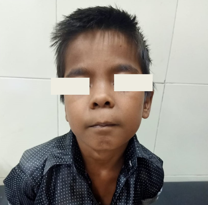
Figure 1: Facial features of child; flat forehead, depressed nasal bridge, low set ear, upturned nose, long philtrum and thin upper lip.
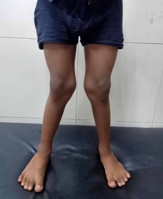
Figure 2: Lower limbs with bilateral genu valgum and sandal gap deformity.
Robinow syndrome is a rare entity in India. It was first described in 1969 as a dwarfing syndrome. It is characterized by short limb dwarfism, deformities in face, head and external genitalia associated with vertebral anomalies (2, 3). It is also called as fetal facies syndrome as the facial features of patients resemble with a fetus. Robinow syndrome has an incidence of 1:500,000 with no gender predilection (4). A few studies have reported higher frequency of Robinow syndrome in some parts of the world especially Turkey and Czechoslovakia (1). On the basis of inheritance both autosomal dominant and recessive forms have been described. The autosomal dominant Robinow syndrome is due to heterozygous mutation in ROR2 gene and WNT5A gene on chromosome 3p whereas the recessive form is due to homozygous mutations localized to chromosome 9q22 (5). Features common in both the dominant and recessive forms are the characteristic facial features, gingival hypertrophy, orodental abnormalities, and maldeveloped genitalia. There is midfacial hypoplasia, short upturned nose with flattening of nasal bridge, broad and prominent forehead, hypertelorism, low set ears, inverted "V" shaped (tented or downturned) upper lip. Both the primary and secondary sets of teeth are present causing malocclusion and overcrowding, often the incisors being exposed due to the maldevelopment of the upper lip. Usually, gum hypertrophy is present from birth. Ankyloglossia and bifid tongue may be associated features. Genital abnormalities are micropenis, cryptorchidism and hypospadias. Hemivertebrae and scoliosis are the distinctive features of recessive form whereas umbilical hernia, supernumerary teeth wide retromolar ridge, alveolar ridge deformation, malocclusion, gingival enlargement, dental crowding, and hypodontia are found mostly in patients with the dominant form (6). Skeletal features with associated genitourinary deformities in the present case suggested strong possibility of Robinow syndrome; however genetic analysis is required for the confirmation and categorization of the syndrome. Genetic testing done antenatally in suspected (especially short stature mothers) would help in early diagnosis of the disease, which can be coupled with effective genetic counseling. Short stature can be well detected by fetal ultrasound evaluation during the routine anomaly scan at 20 weeks gestation (7). Orthopedic management for the skeletal deformities includes postural or surgical correction.
Patton, M.A., and A.R. Afzal. “Robinow Syndrome.” Journal of Medical Genetics, vol. 39, 2002, pp. 305–310.
Soman, C., and A. Lingappa. “Robinow Syndrome: A Rare Case Report and Review of Literature.” International Journal of Clinical Pediatric Dentistry, vol. 8, no. 2, 2015, pp. 149–152.
Hosalkar, H.S., and G.J. Shaw. “Robinow Syndrome.” Journal of Postgraduate Medicine, vol. 48, 2002, pp. 50–51.
Wadia, R.S., D.B. Shirole, and M.S. Dikshit. “Recessively Inherited Costovertebral Segmentation Defect with Mesomelia and Peculiar Facies (Covesdem Syndrome): A New Genetic Entity?” Journal of Medical Genetics, vol. 15, no. 2, 1978, pp. 123–127.
Schwarzer, W., F. Witte, A. Rajab, S. Mundlos, and S. Stricker. “A Gradient of ROR2 Protein Stability and Membrane Localization Confers Brachydactyly Type B or Robinow Syndrome Phenotypes.” Human Molecular Genetics, vol. 18, 2009, pp. 4013–4021.
Beiraghi, S., V. Leon-Salazar, B.E. Larson, M.T. John, M.L. Cunningham, A. Petryk, et al. “Craniofacial and Intraoral Phenotype of Robinow Syndrome Forms.” Clinical Genetics, vol. 80, 2011, pp. 15–24.
Castro, S., E. Peraza, A. Barraza, and M. Zapata. “Prenatal Diagnosis of Robinow Syndrome: A Case Report.” Journal of Clinical Ultrasound, vol. 42, 2014, pp. 297–300.