Contents
Download PDF
pdf Download XML
1927 Views
71 Downloads
Share this article
Research Article | Volume 7 Issue 1 (January-June, 2026) | Pages 1 - 5
Systemic Inflammatory Markers in Patients with Combined Otitis Media and Rhinosinusitis
 ,
 ,
1
Ministry of Health. Karkh Health Directorate, Baghdad, Iraq
2
Ministry of Health, Al-Rusafa Health Directorate, Baghdad, Iraq
Under a Creative Commons license
Open Access
Received
Nov. 3, 2025
Revised
Dec. 9, 2025
Accepted
Jan. 5, 2026
Published
Jan. 28, 2026
Abstract

Background: Otitis media and rhinosinusitis are common inflammatory disorders of the upper airway that frequently coexist due to shared anatomical and immunological pathways. Increasing evidence supports the concept of a unified airway, suggesting that inflammation of the nasal cavity and paranasal sinuses may extend to the middle ear.  Objective: To evaluate systemic inflammatory markers in patients with combined otitis media and rhinosinusitis and compare them with healthy controls. Methods: This hospital-based case–control study was conducted at the Department of Otolaryngology, Al-Kindy Teaching Hospital, Baghdad, Iraq, between March 1, 2025, and December 30, 2026. A total of 140 participants were enrolled, including 70 patients diagnosed with combined otitis media and rhinosinusitis and 70 age- and sex-matched healthy controls. Clinical diagnosis was confirmed through otoscopic examination, nasal endoscopy, and radiological assessment when indicated. Venous blood samples were collected to measure systemic inflammatory markers including white blood cell count (WBC), neutrophil count, lymphocyte count, neutrophil-to-lymphocyte ratio (NLR), platelet count, platelet-to-lymphocyte ratio (PLR), C-reactive protein (CRP), erythrocyte sedimentation rate (ESR), and hemoglobin levels. Results: No significant differences were observed between groups regarding age or sex distribution (p>0.05). Patients with combined OM and RS demonstrated significantly elevated WBC (9.80±2.10 vs. 6.70±1.50 ×10⁹/L), neutrophil count (6.50±1.90 vs. 3.80±1.20 ×10⁹/L), NLR (3.50±1.20 vs. 1.40±0.60), platelet count (320.00±60.00 vs. 260.00±50.00 ×10⁹/L), PLR (160.00±40.00 vs. 95.00±30.00), CRP (12.40±5.60 vs. 3.20±1.80 mg/L), and ESR (28.60±10.20 vs. 12.30±5.40 mm/hr) compared with controls (all p < 0.001). Lymphocyte count was significantly reduced in cases (2.10±0.70 vs. 2.80±0.80 ×10⁹/L, p < 0.001). Hemoglobin was mildly but significantly lower in cases (12.60±1.40 vs. 13.20±1.30 g/dL, p = 0.02). Correlation analysis demonstrated significant associations among inflammatory indices, particularly between NLR and CRP, ESR, and neutrophil count (p<0.001). Conclusion: Combined otitis media and rhinosinusitis are associated with significant systemic inflammatory response. Hematological inflammatory indices such as NLR, PLR, CRP, and ESR may serve as accessible and cost-effective markers reflecting disease burden and inflammatory severity. Incorporating systemic inflammatory markers into clinical evaluation may enhance patient stratification and management strategies.

Keywords
INTRODUCTION

Occurring worldwide, otitis media (OM) and rhinosinusitis (RS) are among the most prevalent inflammatory disorders in otolaryngology practice [1-2]. These conditions have a fundamental effect on quality of life as well as healthcare utilization and resources since they are recurrent and chronic in nature [3]. The "unified airway" theory is increasingly being supported by evidence suggesting a pathiophysiological correlation between inflammatory processes occurring within the nasal cavity, paranasal sinuses, and middle ear [4-5]. The Eustachian tube is a major anatomical and functional link between the nasopharynx and middle ear, permitting bidirectional propagation of inflammatory mediators and pathogens [6-7]. Chronic rhinosinusitis has frequently been associated with Eustachian tube dysfunction and middle ear effusion [8-9]. Similarly, otitis media is often comorbid with sinonasal inflammatory disease, more so in patients with allergic or chronic inflammatory phenotypes [10-11]. Systemic inflammation underpins the common theme in upper airway diseases [12]. These studies may move away from only mucosal inflammation to pick up on some systemic signs of inflammation driving the acute and chronic conditions of ENT [13-14]. Circulatory markers like total WBC (white blood cell) count, NLR, PLR, CRP, and ESR are showing promise as indicators of systemic immune activation [15-18]. Elevated NLR has been associated with chronic rhinosinusitis and associated with increased disease severity and recurrence risk [19-20]. High PLR has also been associated with otitis media with effusion and chronic inflammatory otologic conditions [21-22]. CRP was raised in both acute otitis media infections and bacterial rhinosinusitis because CRP is an index of a systemic acute-phase response [23-24]. It may, in fact, have further implications in the increased inflammatory response seen in patients with concomitant diseases of OM and RS compared to those presenting with solitary ailments [25]. However, limited reports exist on the assessment of these systemic inflammatory markers in this combined phenotype, particularly in adults [26-27]. An appreciation of the systemic inflammatory pattern in this patient group could help in risk stratification and therapeutic decision-making. Therefore, this study is aimed at exploring the systemic inflammatory biomarkers in patients with combined otitis media and rhinosinusitis in comparison to healthy controls and to evaluate their potential significance in clinical practice.

MATERIALS AND METHODS

Patients and Methods

This hospital-based case-control study was conducted at the Department of Otolaryngology, of the Al-Kindy Teaching Hospital, Baghdad, Iraq, during the period between March 1, 2015, and December 30, 2016. The study has been performed by consultants in otolaryngology with specialists in clinical laboratory. The institutional scientific board provided ethical approval following written informed consent secured from each participant before recruitment commenced.


Overall, there were 140 adult participants in the study, divided equally into two groups. The case group was comprised of 70 patients who were diagnosed with otitis media combined with rhinosinusitis, while the control group was made up of 70 healthy people of either sex who were matched for age and without signs of an ENT inflammatory disease on clinical examination. The cases underwent consecutive recruitment over the course of interactions in the ENT outpatient clinic. Controls were selectees at the help of hospital staff and relatives of patients who were included in the study and found free of any otologic or sinonasal pathologies.


Inclusion criteria for the case group included a clinically confirmed diagnosis of otitis media whose diagnosis was based on otoscopic findings of congestion, effusion, and retraction of the tympanic membrane, along with radiological and/or endoscopic confirmation of rhinosinusitis. Recruited cases were patients at least 18 years of age.


Participants that had autoimmune disorders, malignancy, systemic inflammatory disorders, had undergone recent systemic corticosteroid or immune-suppressive therapy, were in systemic infections, or have known hematologic problems were excluded, with the objective of decreasing confounding elements that could affect systemic inflammatory markers.


All patients were assessed by experienced ENT specialists using a standardized protocol, which began with the taking of a detailed medical history focused on the onset and recurrence of symptoms, allergy history, smoking status, and prior treatments received. Otoscopy using a high-definition otoscope was employed to inspect the health status of the middle ear. Nasal endoscopy was performed to visualize mucosal edema, purulent discharge, nasal polyps, and ostiomeatal complex occlusion. When clinically warranted, computed tomography (CT) of the paranasal sinuses was carried out for confirmation of sinus involvement and assessment of disease extent.


Under carefully controlled morning conditions, venous blood samples were taken to ensure harmony in laboratory parameters. Samples were then taken to the central laboratory for processing following established procedures. Inflammatory markers from the systemic bloodstream were measured as total WBC count, absolute neutrophil count, absolute lymphocyte count, NLR, platelet count, platelet-to-lymphocyte ratio (PLR), CRP, ESR, and hemoglobin. CBC was analyzed through automation hematology analyzers, while CRP was assessed through a high-sensitivity immunoturbidimetric means. ESR was measured with the Westergren method.


Continuous variables followed by descriptive statistics are Means±SD, while categorical variables are given as n (%). These authenticated (by the chi-square test) the distribution. Pearson correlation analysis was used for determining the associations between markers. Factors were considered statistically significant when p<0.05.

RESULTS

The study intimated that there was no dissimilarity between the cases and the controls with regards to their age means, which were 38.40 years (SD = 12.60) and 37.90 years (SD = 11.80) respectively, as per Mann-Whitney U test (p = 0.81). This is further deducible from the distribution of male components with cases making 57.14% (40 males) and female components with 54.29% (38 male cases), with a p-Value of 0.72 (Table 1).


Table 1: Demographic Characteristics

VariableCases (n = 70)Controls (n = 70)p-Value
Age (years)38.40±12.6037.90±11.800.81
Male40 (57.14%)38 (54.29%)0.72
Female30 (42.86%)32 (45.71%)-


The study showed significantly elevated total WBC count in cases (9.80±2.10 ×10⁹/L) compared with controls (6.70±1.50 ×10⁹/L), p < 0.001, indicating systemic inflammatory activation (Table 2).


Table 2: White Blood Cell Count

Variable

Cases

Controls

p-Value

WBC (×10⁹/L)

9.80±2.10

6.70±1.50

<0.001


Neutrophil count was significantly higher among cases (6.50±1.90 ×10⁹/L) compared to controls (3.80±1.20 ×10⁹/L), p<0.001 (Table 3).


Table 3: Neutrophil Count

VariableCasesControlsp-Value
Neutrophils (×10⁹/L)6.50±1.903.80±1.20<0.001


Lymphocyte count was significantly reduced in cases (2.10±0.70 ×10⁹/L) compared with controls (2.80±0.80 ×10⁹/L), p<0.001 (Table 4).


Table 4: Lymphocyte Count

Variable

Cases

Controls

p-Value

Lymphocytes (×10⁹/L)

2.10±0.70

2.80±0.80

<0.001


The neutrophil-to-lymphocyte ratio (NLR) was significantly elevated in cases (3.50±1.20) versus controls (1.40±0.60), p<0.001 (Table 5).


Table 5: Neutrophil-to-Lymphocyte Ratio

Variable

Cases

Controls

p-Value

NLR

3.50±1.20

1.40±0.60

<0.001


Platelet count was significantly increased in cases (320.00±60.00 ×10⁹/L) compared to controls (260.00±50.00 ×10⁹/L), p<0.001 (Table 6).


Table 6: Platelet Count

Variable

Cases

Controls

P-Value

Platelets (×10⁹/L)

320.00±60.00

260.00±50.00

<0.001


PLR was significantly higher in cases (160.00±40.00) compared to controls (95.00±30.00), p<0.001 Table 7.


Table 7: Platelet-to-Lymphocyte Ratio

Variable

Cases

Controls

p-Value

PLR

160.00±40.00

95.00±30.00

<0.001


CRP levels were markedly elevated in cases (12.40±5.60 mg/L) versus controls (3.20±1.80 mg/L), p<0.001 Table 8.


Table 8: C-Reactive Protein

Variable

Cases

Controls

p-Value

CRP (mg/L)

12.40±5.60

3.20±1.80

<0.001


ESR was significantly increased in cases (28.60±10.20 mm/hr) compared to controls (12.30±5.40 mm/hr), p<0.001 (Table 9).


Table 9: Erythrocyte Sedimentation Rate

Variable

Cases

Controls

p-Value

ESR (mm/hr)

28.60±10.20

12.30±5.40

<0.001


Hemoglobin was slightly lower in cases (12.60±1.40 g/dL) compared to controls (13.20±1.30 g/dL), with statistical significance (p = 0.02) (Table 10).


Table 10: Hemoglobin Level

Variable

Cases

Controls

P-Value

Hemoglobin (g/dL)

12.60±1.40

13.20±1.30

0.02


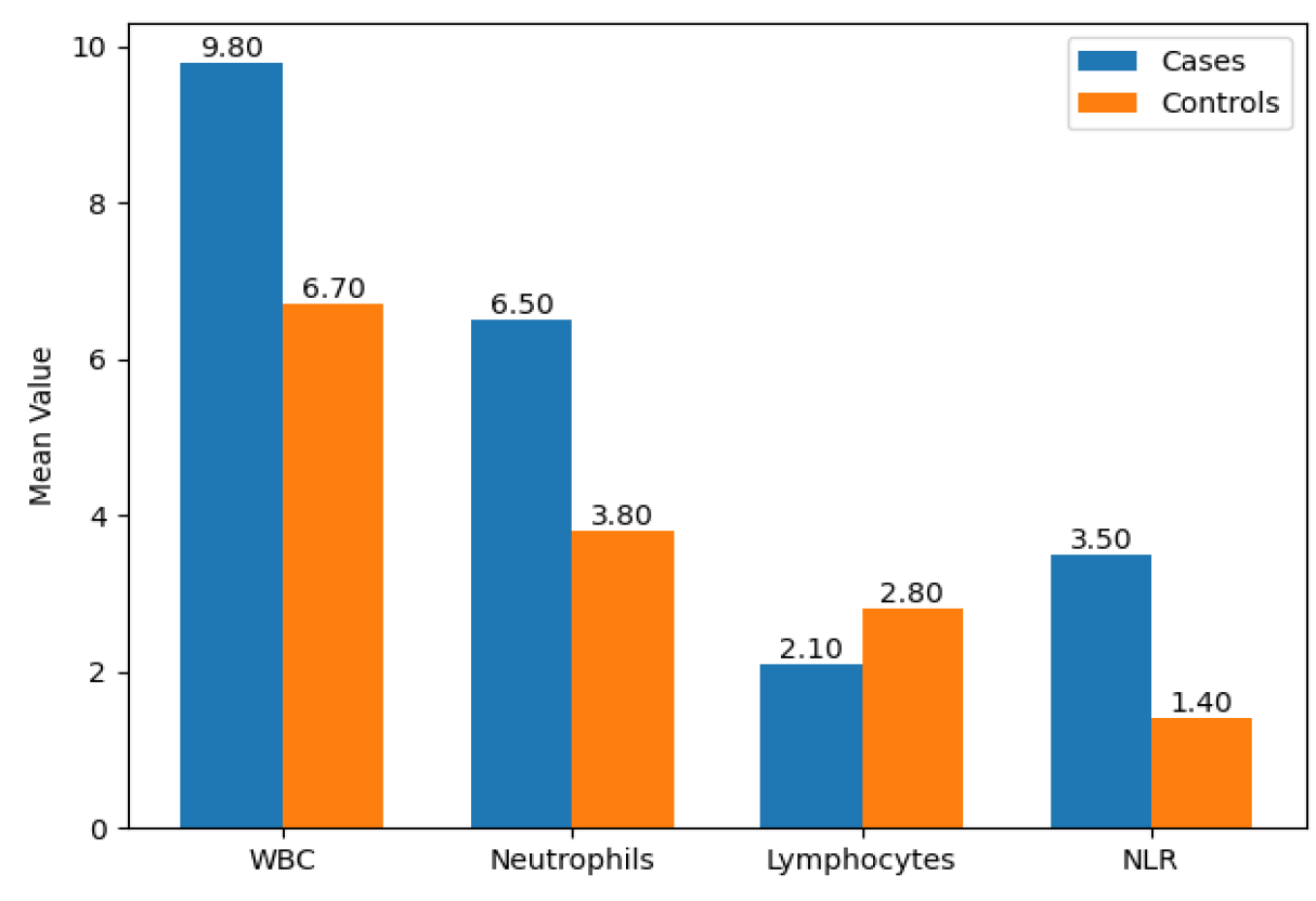
Pearson correlation analysis demonstrated that lymphocyte count was significantly negatively correlated with NLR (r = −0.68, p < 0.001). Platelet count showed a moderate positive correlation with PLR (r = 0.61, p<0.001). CRP and ESR demonstrated strong positive correlations with NLR (r = 0.59 and r = 0.55, respectively; both p<0.001). Hemoglobin showed a mild negative correlation with CRP (r = −0.29, p = 0.01). These findings confirm that elevated systemic inflammatory markers are interrelated and reflect overall inflammatory burden in patients with combined otitis media and rhinosinusitis Table 11 (Figure 1).


Table 11: Correlation between Systemic Inflammatory Markers in Cases (n = 70)

Variable

NLR (r)

PLR (r)

CRP (r)

ESR (r)

p-Value

Lymphocytes (×10⁹/L)

−0.68

−0.54

−0.49

−0.46

<0.001

Neutrophils (×10⁹/L)

0.72

0.51

0.58

0.53

<0.001

Platelets (×10⁹/L)

0.44

0.61

0.47

0.42

<0.001

CRP (mg/L)

0.59

0.52

0.63

<0.001

ESR (mm/hr)

0.55

0.49

0.63

<0.001

Hemoglobin (g/dL)

−0.31

−0.27

−0.29

−0.25

0.01


Figure 1: Comparison of Systemic Inflammatory Markers between Cases and Controls

DISCUSSION

This study underscores elevated systemic inflammatory markers in patients suffering from combined otitis media and rhinosinusitis compared to controls. These escalated values of WBC, absolute neutrophils, neutrophil-lymphocyte ratio, platelet-lymphocyte ratio, C-reactive protein (CRP), and erythrocyte sedimentation rate (ESR) suggest that systemic immune activation is present with current concurrent inflammation of the middle ear and sinonasal cavity. The concept of a united airway could explain the results of our study [4-5]. Cytokines released in the nose and nasal tissues might reach the middle ears via continuity of the mucosa and thereby the systemic circulation and may enhance inflammation in the middle ear [6,28]. Earlier studies have also suggested that middle ear effusion and Eustachian tube dysfunction have heights in patients with chronic rhinosinusitis [8,9,29]. Conclusively, the finding of significantly raised WBC and neutrophils (p<0.001) fits with earlier reports where neutrophils get to behave in predominance in bacterial rhinosinusitis and otitis media [12-13]. Neutrophil-inspired tissue harm can be linked to reactive oxygen species and proteases [30]. The increase in NLR is consistent with other studies done which also describe the same in various ENT diseases [15,19-20]. Elevations in PLR are consistent with an individual-level increase in platelet activation, enhancing the supposition of inflammasome and endothelial disgracing [16,21]. Platelets have the capability to recruit immune cells and excessively release cytokines for the so-called reinforcement of inflammatory mechanisms in upper airway diseases [31]. CRP and ESR were markedly elevated in cases (p < 0.001) in line with systemic inflammation and acute-phase response [17,23]. Increased cartilage-binding CRP has been associated with bacterial cleansing capacity and disease extent in cases of otitis media and rhinosinusitis [24,32]. Raising ESR portrays a long-standing burden of inflammation [18]. In declined hemoglobin values (though discrete), further evidence may exist quantifying restrictions to erythropoiesis by inflammation in a variety of chronic inflammatory states [33]. Inflammatory cytokine-rich chronic sinonasal inflammation links hemorrhage-systemic cytokine mediated hematological changes [34]. The combined spectrum of OM and RS may have a more serious inflammatory phenotype when compared with singular disease [25,27]. What is found in the literature regarding proinflammatory mediators in ME effusion implies that raised ILs and tumor necrosis factors are seen with concurrent sinonasal disease [35]. Clinically, systemic inflammatory markers, if abnormal, can also act as adjunctive tools in identifying patients at risk for persistent/recurrent disease. The recognition of systemic inflammation is vital for the initiation of intensified close follow-up or more aggressive therapy. The study is strong as it leverages the case–control design and universally useful laboratory evaluations, yet it is subject to narrowness due to being a single-center study, with the lack of cytokine profiling. Future works focused on the molecular markers of inflammation would be welcomed.

REFERENCE
  1. Marple, B.F. “Allergic rhinitis and inflammatory airway disease: Interactions within the unified airspace.” American Journal of Rhinology & Allergy, vol. 24, no. 4, 2010, pp. 249–254.

  2. Nokso-Koivisto, J. et al. “Otitis media: Interactions between host and environment, immune and inflammatory responses.” International Journal of Pediatric Otorhinolaryngology, vol. 178, 2024, pp. 111510.

  3. Corren, J. “The link between allergic rhinitis and asthma, otitis media, sinusitis, and nasal polyps.” Radiologic Clinics of North America, vol. 38, no. 6, 2000, pp. 1257–1266.

  4. Gentile, D.A. et al. “Otitis media and sinusitis.” Allergy Frontiers: Clinical Manifestations, edited by R.Pawankar et al., Springer, 2009, pp. 457–472.

  5. Van Cauwenberge, P. et al. “Glucocorticosteroids in allergic inflammation: Clinical benefits in allergic rhinitis, rhinosinusitis, and otitis media.” Immunology and Allergy Clinics of North America, vol. 25, no. 2, 2005, pp. 321–342.

  6. Chang, W. and X. Zhou. “Effects of combined balloon dilatation and tympanic membrane catheterization on Eustachian tube function and hearing recovery in secretory otitis media.” Medical Science Monitor, vol. 31, 2025, pp. e942310.

  7. Kim, S.Y. et al. “Bidirectional association between asthma and otitis media in children.” Allergy, Asthma & Clinical Immunology, vol. 17, 2021, pp. 77.

  8. Parietti-Winkler, C. et al. “Otitis media with effusion as a marker of the inflammatory process associated with nasal polyposis.” European Archives of Oto-Rhino-Laryngology, vol. 266, no. 3, 2009, pp. 339–344.

  9. Iino, Y. “Eosinophilic otitis media: State-of-the-art diagnosis and treatment.” Auris Nasus Larynx, vol. 50, no. 4, 2023, pp. 473–480.

  10. Ciprandi, G. et al. “Allergy and otitis media in clinical practice.” Current Allergy and Asthma Reports, vol. 20, no. 7, 2020, pp. 33.

  11. Vlastos, I.M. et al. “Is allergic rhinitis related to otitis media with effusion in adults and children?” Cells, vol. 14, no. 2, 2025, pp. 198.

  12. Juhn, S.K. et al. “The role of inflammatory mediators in the pathogenesis of otitis media and sequelae.” Clinical and Experimental Otorhinolaryngology, vol. 1, no. 3, 2008, pp. 117–138.

  13. Hurst, J.R. et al. “Relationships among bacteria, upper airway, lower airway, and systemic inflammation in COPD.” Chest, vol. 127, no. 4, 2005, pp. 1219–1226.

  14. Chkhaidze, I. et al. “Systemic inflammatory responses in patients with acute otitis media and the impact of treatment.” Georgian Medical News, no. 152, 2007, pp. 37–42.

  15. Kaminer, B.M. et al. “Neutrophil-to-lymphocyte ratio as biomarker in ENT infections.” Clinical Otolaryngology, vol. 49, no. 1, 2024, pp. 112–118.

  16. Tuna, B. and G.Tüzemen. “The role of blood inflammatory parameters in otitis media with effusion in children with obstructive adenoid hypertrophy.” Genel Tıp Dergisi, vol. 35, no. 2, 2025, pp. 145–152.

  17. Arikan, T.A. and M.Kelles. “Plasma selenium and cadmium levels in patients with chronic otitis media and relation to inflammation markers.” Biological Trace Element Research, vol. 189, no. 2, 2019, pp. 329–336.

  18. Sakovets, O.P. et al. “Immunohematological indices as markers of inflammation in children with acute otitis media.” Clinical and Experimental Otorhinolaryngology, vol. 18, no. 1, 2025, pp. 45–52.

  19. Yeniceri, A., and M.Cayonu. “Outcomes of inflammatory markers in the diagnosis of complicated rhinosinusitis in children.” European Archives of Oto-Rhino-Laryngology, vol. 282, no. 3, 2025, pp. 715–722.

  20. Yang, J. et al. “Correlation analysis of serum inflammatory markers in secretory otitis media.” Journal of Medical Sciences, vol. 46, no. 1, 2026, pp. 22–29.

  21. Almutairi, A.A. et al. “Blood inflammatory markers as predictors in otitis media with effusion.” Medicina, vol. 61, no. 1, 2025, pp. 74.

  22. Esu, Y. et al. “Periostin in middle ear mucosa according to eosinophilic otitis media severity.” Auris Nasus Larynx, vol. 47, no. 6, 2020, pp. 1015–1021.

  23. Manole, F. et al. “Biomarker analysis in upper respiratory tract infections.” Pediatric Reports, vol. 16, no. 2, 2024, pp. 245–256.

  24. Sarı, N. et al. “Role of oxidative stress in serous otitis media and myringosclerosis.” Bratislava Medical Journal, vol. 126, no. 4, 2025, pp. 245–251.

  25. Yoshida, N. “Intractable otitis media – Pathogenesis and treatment.” Auris Nasus Larynx, vol. 50, no. 5, 2023, pp. 650–657.

  26. Peric, A. and D.Djeric. “Immunological mechanisms of sensorineural hearing impairment in chronic rhinosinusitis.” Immunologic Investigations, vol. 54, no. 3, 2025, pp. 412–424.

  27. Li, Y. et al. “Eosinophilic otitis media treated with dupilumab: Case report and review.” Acta Oto-Laryngologica Case Reports, vol. 10, no. 1, 2025, pp. 56–62.

  28. Manciula, L.G. et al. “Biofilms and inflammation in chronic rhinosinusitis.” Clujul Medical, vol. 93, no. 2, 2020, pp. 180–186.

  29. Lechien, J.R. et al. “Association between laryngopharyngeal reflux and otitis media: Systematic review.” Otology & Neurotology, vol. 42, no. 5, 2021, pp. 680–689.

  30. Brown, K.L. et al. “Complexities of neutrophil interactions in inflammation.” Journal of Immunology, vol. 189, no. 2, 2012, pp. 533–539.

  31. Koupenova, M. et al. “Circulating platelets as mediators of immunity and inflammation.” Circulation Research, vol. 122, no. 2, 2018, pp. 337–351.

  32. Rosenfeld, R.M. et al. “Clinical practice guideline (update): Adult sinusitis.” Otolaryngology–Head and Neck Surgery, vol. 152, no. 2 Suppl., 2015, pp. S1–S39.

  33. Weiss, G. and L.T.Goodnough. “Anemia of chronic disease.” New England Journal of Medicine, vol. 352, no. 10, 2005, pp. 1011–1023.

  34. Fokkens, W.J. et al. “European position paper on rhinosinusitis and nasal polyps 2020.” Rhinology, vol. 58, suppl. S29, 2020, pp. 1–464.

  35. Suikkila, A. et al. “Inflammatory cytokines in middle ear effusion of patients with asthma and chronic rhinosinusitis.” Otology & Neurotology, vol. 45, no. 3, 2024, pp. e233–e240.


License
CC BY-NC-ND
Creative Commons Attribution-NonCommercial-NoDerivatives 4.0 International License
Systemic Inflammatory Markers in Patients with Combined Otitis Media and Rhinosinusitis © 2026 by Mohammed Qasim Mohammed, Watheq A. Mohammed Ali, Abdulkareem Hussien Dabi licensed under CC BY-NC-ND 4.0
All papers should be submitted electronically. All submitted manuscripts must be original work that is not under submission at another journal or under consideration for publication in another form, such as a monograph or chapter of a book. Authors of submitted papers are obligated not to submit their paper for publication elsewhere until an editorial decision is rendered on their submission. Further, authors of accepted papers are prohibited from publishing the results in other publications that appear before the paper is published in the Journal unless they receive approval for doing so from the Editor-In-Chief.
Himalayan Journal of Medicine and Surgery open access articles are licensed under a Creative Commons Attribution-Share A like 4.0 International License. This license lets the audience to give appropriate credit, provide a link to the license, and indicate if changes were made and if they remix, transform, or build upon the material, they must distribute contributions under the same license as the original.
Recommended Articles
Research Article
Primordial Prevention of Coronary Artery Diseases
...
Published: 26/07/2026
Download PDF
Research Article
Impact of Gut-Liver Axis: Hepatic Biochemical and Metabolic Changes Associated with Chronic Gastritis in Iraqi Patients with Helicobacter Pylori
Published: 20/02/2026
Download PDF
Research Article
Effect of Land Degradation on Livelihood
Published: 04/01/2024
Download PDF
Research Article
Awareness and Practices among Professional Healthcare Workers towards COVID-19 in Iraq
...
Published: 30/03/2024
Download PDF
Flowbite Logo
Najmal Complex,
Opposite Farwaniya,
Kuwait.
Email: support@himjournals.com

Useful Links
Order Hard Copy
Privacy policy
Terms and Conditions
Refund Policy
Others
About Us
Team Members
Contact Us
Online Payments
Join as Editor
Join as Reviewer
Subscribe to our Newsletter
Follow us
MOST SEARCHED KEYWORDS
scientific journal
 | 
business journal
 | 
medical journals
 | 
Scientific Journals
 | 
Academic Publisher
 | 
Peer-reviewed Journals
 | 
Open Access Journals
 | 
Impact Factor
 | 
Indexing Services
 | 
Journal Citation Reports
 | 
Publication Process
 | 
Impact factor of journals
 | 
Finding reputable journals for publication
 | 
Submitting a manuscript for publication
 | 
Copyright and licensing of published papers
 | 
Writing an abstract for a research paper
 | 
Manuscript formatting guidelines
 | 
Promoting published research
 | 
Publication in high-impact journals
Copyright © Himalayan Journals . All Rights Reserved.